 Movie
Movie Controller


+ Open data

- Basic information
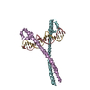
| Entry | Database: PDB / ID: 3a5t | ||||||
|---|---|---|---|---|---|---|---|
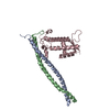
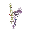
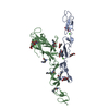
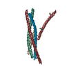
| Title | Crystal structure of MafG-DNA complex | ||||||
 Components Components |
| ||||||
 Keywords Keywords | TRANSCRIPTION REGULATOR/DNA /  PROTEIN-DNA COMPLEX / BZIP FACTOR / Acetylation / DNA-binding / Isopeptide bond / Nucleus / Repressor / Transcription / Transcription regulation / Ubl conjugation / TRANSCRIPTION REGULATOR-DNA COMPLEX PROTEIN-DNA COMPLEX / BZIP FACTOR / Acetylation / DNA-binding / Isopeptide bond / Nucleus / Repressor / Transcription / Transcription regulation / Ubl conjugation / TRANSCRIPTION REGULATOR-DNA COMPLEX | ||||||
| Function / homology |  Function and homology information Function and homology informationFactors involved in megakaryocyte development and platelet production / regulation of epidermal cell differentiation / regulation of cellular pH / adult behavior / RNA polymerase II transcription regulator complex / sequence-specific double-stranded DNA binding / regulation of cell population proliferation / DNA-binding transcription activator activity, RNA polymerase II-specific / in utero embryonic development / transcription regulator complex ...Factors involved in megakaryocyte development and platelet production / regulation of epidermal cell differentiation / regulation of cellular pH / adult behavior / RNA polymerase II transcription regulator complex / sequence-specific double-stranded DNA binding / regulation of cell population proliferation / DNA-binding transcription activator activity, RNA polymerase II-specific / in utero embryonic development / transcription regulator complex / DNA-binding transcription factor activity, RNA polymerase II-specific / RNA polymerase II cis-regulatory region sequence-specific DNA binding / positive regulation of gene expression / regulation of transcription by RNA polymerase II / positive regulation of transcription by RNA polymerase II / DNA binding / identical protein binding / nucleusSimilarity search - Function | ||||||
| Biological species |  Mus musculus (house mouse) Mus musculus (house mouse)synthetic construct (others) | ||||||
| Method | X-RAY DIFFRACTION / SYNCHROTRON / MAD / Resolution: 2.8 Å | ||||||
 Authors Authors | Kurokawa, H. / Motohashi, H. / Sueno, S. / Kimura, M. / Takagawa, H. / Kanno, Y. / Yamamoto, M. / Tanaka, T. | ||||||
 Citation Citation | Journal: Mol.Cell.Biol. / Year: 2009 Title: Structural Basis of Alternative DNA Recognition by Maf Transcription Factors Authors: Kurokawa, H. / Motohashi, H. / Sueno, S. / Kimura, M. / Takagawa, H. / Kanno, Y. / Yamamoto, M. / Tanaka, T. | ||||||
| History |
| ||||||
| Remark 650 | HELIX DETERMINATION METHOD: AUTHOR DETERMINED |
- Structure visualization
| Structure viewer | Molecule: MolmilJmol/JSmol |
|---|
- Downloads & links
-Download
| PDBx/mmCIF format | 3a5t.cif.gz | 64.1 KB | Display | PDBx/mmCIF format |
|---|---|---|---|---|
| PDB format | pdb3a5t.ent.gz | 49.2 KB | Display | PDB format |
| PDBx/mmJSON format | 3a5t.json.gz | Tree view | PDBx/mmJSON format | |
| Others |  Other downloads Other downloads |
-Validation report
| Arichive directory | https://data.pdbj.org/pub/pdb/validation_reports/a5/3a5tftp://data.pdbj.org/pub/pdb/validation_reports/a5/3a5t | HTTPS FTP |
|---|
-Related structure data
| Similar structure data |
|---|

-Links
 PDBj
PDBj


- Assembly
| Deposited unit | 
| ||||||||
|---|---|---|---|---|---|---|---|---|---|
| 1 |
| ||||||||
| Unit cell |
|
-Components
| #1: Protein | Mass: 12521.845 Da / Num. of mol.: 2 / Fragment: BINDING DOMAIN, RESIDUES 21-123 Source method: isolated from a genetically manipulated source Source: (gene. exp.) Mus musculus (house mouse) / Gene: Mafg / Plasmid: pET15B / Production host:  Escherichia coli (E. coli) / Strain (production host): BL21(DE3)-CodonPlus-RIL / References: UniProt: O54790 Escherichia coli (E. coli) / Strain (production host): BL21(DE3)-CodonPlus-RIL / References: UniProt: O54790#2: DNA chain | | Mass: 4593.998 Da / Num. of mol.: 1 / Source method: obtained synthetically / Source: (synth.) synthetic construct (others) #3: DNA chain | | Mass: 4584.985 Da / Num. of mol.: 1 / Source method: obtained synthetically / Source: (synth.) synthetic construct (others) #4: Chemical | ChemComp-MG / |   Mass: 24.305 Da / Num. of mol.: 1 / Source method: obtained synthetically / Formula: Mg Mass: 24.305 Da / Num. of mol.: 1 / Source method: obtained synthetically / Formula: Mg#5: Water | ChemComp-HOH / | Water Mass: 18.015 Da / Num. of mol.: 83 / Source method: isolated from a natural source / Formula: H2O Mass: 18.015 Da / Num. of mol.: 83 / Source method: isolated from a natural source / Formula: H2O |
|---|
-Experimental details
-Experiment
| Experiment | Method: X-RAY DIFFRACTION / Number of used crystals: 1 |
|---|
- Sample preparation
| Crystal | Density Matthews: 6.288576 Å3/Da / Density % sol: 80.44072 % |
|---|---|
| Crystal grow | Temperature: 298 K / Method: vapor diffusion, hanging drop / pH: 5 Details: 0.1M sodium acetate, 40mM magnesium chloride hexahydrate, 8% 2-methyl-2,4-pentandiol, 4mM Tris(2-carboxyethyl)phosphine, pH 5.0, VAPOR DIFFUSION, HANGING DROP, temperature 298K |
-Data collection
| Diffraction | Mean temperature: 100 K | ||||||||||||
|---|---|---|---|---|---|---|---|---|---|---|---|---|---|
| Diffraction source | Source: SYNCHROTRON / Site: Photon Factory  / Beamline: BL-17A / Wavelength: 0.97939, 0.97898, 0.96416 / Beamline: BL-17A / Wavelength: 0.97939, 0.97898, 0.96416 | ||||||||||||
| Detector | Type: ADSC QUANTUM 4r / Detector: CCD / Date: Dec 14, 2006 / Details: mirrors | ||||||||||||
| Radiation | Monochromator: Numerical link type Si(111) double crystal / Protocol: MAD / Monochromatic (M) / Laue (L): M / Scattering type: x-ray | ||||||||||||
| Radiation wavelength |
| ||||||||||||
| Reflection | Resolution: 2.8→36.08 Å / Num. obs: 22101 / % possible obs: 98.9 % / Redundancy: 13 % / Biso Wilson estimate: -0.8 Å2 / Rmerge(I) obs: 0.072 / Net I/σ(I): 56.77 / Num. measured all: 284413 | ||||||||||||
| Reflection shell | Resolution: 2.8→2.9 Å / Redundancy: 10.2 % / Rmerge(I) obs: 0.192 / Mean I/σ(I) obs: 11.91 / Num. unique all: 2016 / % possible all: 92.6 |
- Processing
| Software |
| ||||||||||||||||||||||||||||||||||||||||||||||||||||||||||||||||||||||||||||||||
|---|---|---|---|---|---|---|---|---|---|---|---|---|---|---|---|---|---|---|---|---|---|---|---|---|---|---|---|---|---|---|---|---|---|---|---|---|---|---|---|---|---|---|---|---|---|---|---|---|---|---|---|---|---|---|---|---|---|---|---|---|---|---|---|---|---|---|---|---|---|---|---|---|---|---|---|---|---|---|---|---|---|
| Refinement | Method to determine structure: MAD / Resolution: 2.8→36.08 Å / Rfactor Rfree error: 0.006 / Data cutoff high absF: 59212.69 / Data cutoff low absF: 0 / Isotropic thermal model: RESTRAINED / Cross valid method: THROUGHOUT / σ(F): 0 / Stereochemistry target values: Engh & Huber / Details: BULK SOLVENT MODEL USED
| ||||||||||||||||||||||||||||||||||||||||||||||||||||||||||||||||||||||||||||||||
| Solvent computation | Solvent model: FLAT MODEL / Bsol: 17.7589 Å2 / ksol: 0.3 e/Å3 | ||||||||||||||||||||||||||||||||||||||||||||||||||||||||||||||||||||||||||||||||
| Displacement parameters | Biso mean: 73.4 Å2
| ||||||||||||||||||||||||||||||||||||||||||||||||||||||||||||||||||||||||||||||||
| Refine analyze |
| ||||||||||||||||||||||||||||||||||||||||||||||||||||||||||||||||||||||||||||||||
| Refinement step | Cycle: LAST / Resolution: 2.8→36.08 Å
| ||||||||||||||||||||||||||||||||||||||||||||||||||||||||||||||||||||||||||||||||
| Refine LS restraints |
| ||||||||||||||||||||||||||||||||||||||||||||||||||||||||||||||||||||||||||||||||
| LS refinement shell | Resolution: 2.8→2.98 Å / Rfactor Rfree error: 0.026 / Total num. of bins used: 6
|
