 Movie
Movie Controller

[English] 日本語
 Yorodumi
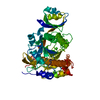
- PDB-3a3p: Crystal structure of complex between E201A/SA-subtilisin and Tk-p... -
+ Open data

- Basic information
| Entry | Database: PDB / ID: 3a3p | ||||||
|---|---|---|---|---|---|---|---|
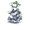
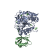
| Title | Crystal structure of complex between E201A/SA-subtilisin and Tk-propeptide | ||||||
 Components Components | (Tk-subtilisin) x 2 | ||||||
 Keywords Keywords |  HYDROLASE / subtilisin / propeptide / Thermococcus kodakaraensis / Protease / Secreted / Serine protease / Zymogen HYDROLASE / subtilisin / propeptide / Thermococcus kodakaraensis / Protease / Secreted / Serine protease / Zymogen | ||||||
| Function / homology |  Function and homology informationHydrolases; Acting on peptide bonds (peptidases); Serine endopeptidases / serine-type endopeptidase activity / proteolysis / extracellular region / identical protein binding Function and homology informationHydrolases; Acting on peptide bonds (peptidases); Serine endopeptidases / serine-type endopeptidase activity / proteolysis / extracellular region / identical protein bindingSimilarity search - Function | ||||||
| Biological species |   Thermococcus kodakarensis (archaea) Thermococcus kodakarensis (archaea) | ||||||
| Method | X-RAY DIFFRACTION / SYNCHROTRON / MOLECULAR REPLACEMENT / Resolution: 1.9 Å | ||||||
 Authors Authors | Tanaka, S. / Matsumura, H. / Koga, Y. / Takano, K. / Kanaya, S. | ||||||
 Citation Citation | Journal: J.Mol.Biol. / Year: 2009 Title: Identification of the interactions critical for propeptide-catalyzed folding of Tk-subtilisin Authors: Tanaka, S. / Matsumura, H. / Koga, Y. / Takano, K. / Kanaya, S. | ||||||
| History |
|
- Structure visualization
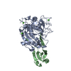
| Structure viewer | Molecule: MolmilJmol/JSmol |
|---|
- Downloads & links
-Download
| PDBx/mmCIF format | 3a3p.cif.gz | 92.3 KB | Display | PDBx/mmCIF format |
|---|---|---|---|---|
| PDB format | pdb3a3p.ent.gz | 67.4 KB | Display | PDB format |
| PDBx/mmJSON format | 3a3p.json.gz | Tree view | PDBx/mmJSON format | |
| Others |  Other downloads Other downloads |
-Validation report
| Arichive directory | https://data.pdbj.org/pub/pdb/validation_reports/a3/3a3pftp://data.pdbj.org/pub/pdb/validation_reports/a3/3a3p | HTTPS FTP |
|---|
-Related structure data
| Related structure data |  3a3nC 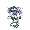 3a3oC  2z30S S: Starting model for refinement C: citing same article ( |
|---|---|
| Similar structure data |


-Links
 PDBj
PDBj

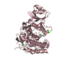
- Assembly
| Deposited unit | 
| ||||||||
|---|---|---|---|---|---|---|---|---|---|
| 1 |
| ||||||||
| Unit cell |
|
-Components
| #1: Protein | Mass: 33839.523 Da / Num. of mol.: 1 / Fragment: Residue in UNP 94-422 / Mutation: E201A, S324A Source method: isolated from a genetically manipulated source Source: (gene. exp.) Thermococcus kodakarensis (archaea) / Plasmid: pET25b / Production host:  Escherichia coli BL21(DE3) (bacteria) / Strain (production host): BL21(DE3) Escherichia coli BL21(DE3) (bacteria) / Strain (production host): BL21(DE3)References: UniProt: P58502, Hydrolases; Acting on peptide bonds (peptidases); Serine endopeptidases | ||||
|---|---|---|---|---|---|
| #2: Protein | Mass: 7512.829 Da / Num. of mol.: 1 / Fragment: Tk-propeptide, Residue in UNP 25-93 Source method: isolated from a genetically manipulated source Source: (gene. exp.) Thermococcus kodakarensis (archaea) / Plasmid: pET25b / Production host: Escherichia coli BL21(DE3) (bacteria) / Strain (production host): BL21(DE3) / References: UniProt: P58502 | ||||
| #3: Chemical | ChemComp-CA /   Mass: 40.078 Da / Num. of mol.: 7 / Source method: obtained synthetically / Formula: Ca Mass: 40.078 Da / Num. of mol.: 7 / Source method: obtained synthetically / Formula: Ca#4: Chemical | ChemComp-ZN / |   Mass: 65.409 Da / Num. of mol.: 1 / Source method: obtained synthetically / Formula: Zn Mass: 65.409 Da / Num. of mol.: 1 / Source method: obtained synthetically / Formula: Zn#5: Water | ChemComp-HOH / | Water Mass: 18.015 Da / Num. of mol.: 236 / Source method: isolated from a natural source / Formula: H2O Mass: 18.015 Da / Num. of mol.: 236 / Source method: isolated from a natural source / Formula: H2O |
-Experimental details
-Experiment
| Experiment | Method: X-RAY DIFFRACTION / Number of used crystals: 1 |
|---|
- Sample preparation
| Crystal | Density Matthews: 1.95 Å3/Da / Density % sol: 36.81 % |
|---|---|
| Crystal grow | Temperature: 293 K / Method: vapor diffusion, sitting drop / pH: 6.5 Details: 0.1M Sodium Cacodylate, 0.2M Zinc Acetate, 10%(v/v) isopropanol, pH 6.5, VAPOR DIFFUSION, SITTING DROP, temperature 293K |
-Data collection
| Diffraction | Mean temperature: 100 K |
|---|---|
| Diffraction source | Source: SYNCHROTRON / Site: SPring-8  / Beamline: BL38B1 / Wavelength: 1 Å / Beamline: BL38B1 / Wavelength: 1 Å |
| Detector | Type: RIGAKU JUPITER 210 / Detector: CCD / Date: Oct 22, 2008 |
| Radiation | Protocol: SINGLE WAVELENGTH / Monochromatic (M) / Laue (L): M / Scattering type: x-ray |
| Radiation wavelength | Wavelength: 1 Å / Relative weight: 1 |
| Reflection | Resolution: 1.9→50 Å / Num. obs: 26027 / % possible obs: 98.9 % / Rmerge(I) obs: 0.186 / Net I/σ(I): 12.8 |
| Reflection shell | Resolution: 1.9→1.97 Å / Rmerge(I) obs: 0.571 / Mean I/σ(I) obs: 1.8 / % possible all: 99.8 |
- Processing
| Software |
| ||||||||||||||||||||||||||||||||||||||||||||||||||||||||||||||||||||||||||||||||||||||||||||||||||||
|---|---|---|---|---|---|---|---|---|---|---|---|---|---|---|---|---|---|---|---|---|---|---|---|---|---|---|---|---|---|---|---|---|---|---|---|---|---|---|---|---|---|---|---|---|---|---|---|---|---|---|---|---|---|---|---|---|---|---|---|---|---|---|---|---|---|---|---|---|---|---|---|---|---|---|---|---|---|---|---|---|---|---|---|---|---|---|---|---|---|---|---|---|---|---|---|---|---|---|---|---|---|
| Refinement | Method to determine structure: MOLECULAR REPLACEMENT Starting model: 2Z30 Resolution: 1.9→26.98 Å / Cor.coef. Fo:Fc: 0.959 / Cor.coef. Fo:Fc free: 0.941 / SU B: 3.29 / SU ML: 0.098 / Isotropic thermal model: RESTRAINED / Cross valid method: THROUGHOUT / ESU R: 0.165 / ESU R Free: 0.148 / Stereochemistry target values: MAXIMUM LIKELIHOOD / Details: HYDROGENS HAVE BEEN ADDED IN THE RIDING POSITIONS
| ||||||||||||||||||||||||||||||||||||||||||||||||||||||||||||||||||||||||||||||||||||||||||||||||||||
| Solvent computation | Ion probe radii: 0.8 Å / Shrinkage radii: 0.8 Å / VDW probe radii: 1.2 Å / Solvent model: MASK | ||||||||||||||||||||||||||||||||||||||||||||||||||||||||||||||||||||||||||||||||||||||||||||||||||||
| Displacement parameters | Biso mean: 26.474 Å2
| ||||||||||||||||||||||||||||||||||||||||||||||||||||||||||||||||||||||||||||||||||||||||||||||||||||
| Refinement step | Cycle: LAST / Resolution: 1.9→26.98 Å
| ||||||||||||||||||||||||||||||||||||||||||||||||||||||||||||||||||||||||||||||||||||||||||||||||||||
| Refine LS restraints |
| ||||||||||||||||||||||||||||||||||||||||||||||||||||||||||||||||||||||||||||||||||||||||||||||||||||
| LS refinement shell | Resolution: 1.899→1.948 Å / Total num. of bins used: 20
|
