 Movie
Movie Controller


+ Open data

- Basic information
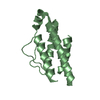
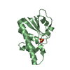
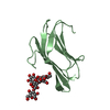
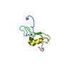
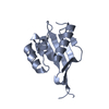
| Entry | Database: PDB / ID: 3a0s | ||||||
|---|---|---|---|---|---|---|---|
| Title | PAS domain of histidine kinase ThkA (TM1359) | ||||||
 Components Components | Sensor protein | ||||||
 Keywords Keywords | TRANSFERASE / PAS-fold / Kinase / Phosphoprotein / Two-component regulatory system | ||||||
| Function / homology |  Function and homology informationhistidine kinase / phosphorelay sensor kinase activity / regulation of DNA-templated transcription / ATP binding Function and homology informationhistidine kinase / phosphorelay sensor kinase activity / regulation of DNA-templated transcription / ATP bindingSimilarity search - Function | ||||||
| Biological species |   Thermotoga maritima (bacteria) Thermotoga maritima (bacteria) | ||||||
| Method | X-RAY DIFFRACTION / SYNCHROTRON / MOLECULAR REPLACEMENT / Resolution: 1.47 Å | ||||||
 Authors Authors | Yamada, S. / Sugimoto, H. / Kobayashi, M. / Ohno, A. / Nakamura, H. / Shiro, Y. | ||||||
 Citation Citation | Journal: Structure / Year: 2009 Title: Structure of PAS-linked histidine kinase and the response regulator complex Authors: Yamada, S. / Sugimoto, H. / Kobayashi, M. / Ohno, A. / Nakamura, H. / Shiro, Y. | ||||||
| History |
|
- Structure visualization
| Structure viewer | Molecule: MolmilJmol/JSmol |
|---|
- Downloads & links
-Download
| PDBx/mmCIF format | 3a0s.cif.gz | 37.1 KB | Display | PDBx/mmCIF format |
|---|---|---|---|---|
| PDB format | pdb3a0s.ent.gz | 25 KB | Display | PDB format |
| PDBx/mmJSON format | 3a0s.json.gz | Tree view | PDBx/mmJSON format | |
| Others |  Other downloads Other downloads |
-Validation report
| Arichive directory | https://data.pdbj.org/pub/pdb/validation_reports/a0/3a0sftp://data.pdbj.org/pub/pdb/validation_reports/a0/3a0s | HTTPS FTP |
|---|
-Related structure data
| Related structure data |  3a0rC 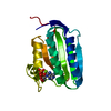 3a0tC  3a0uC  3a0vSC  3a0wC 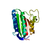 3a0xC 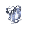 3a0yC 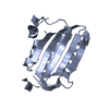 3a0zC 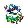 3a10C C: citing same article ( S: Starting model for refinement |
|---|---|
| Similar structure data |


-Links
 PDBj
PDBj


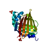
- Assembly
| Deposited unit | 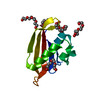
| ||||||||
|---|---|---|---|---|---|---|---|---|---|
| 1 |
| ||||||||
| Unit cell |
|
-Components
| #1: Protein | / Histidine kinase ThkA Mass: 11219.812 Da / Num. of mol.: 1 / Fragment: PAS domain Source method: isolated from a genetically manipulated source Source: (gene. exp.) Thermotoga maritima (bacteria) / Gene: TM_1359 / Plasmid: pRSETA / Production host: Escherichia coli BL21(DE3) (bacteria) / Strain (production host): BL21(DE3) CodonPlus RIL / References: UniProt: Q9X180, histidine kinase | ||||||
|---|---|---|---|---|---|---|---|
| #2: Chemical | ChemComp-PG4 / Polyethylene glycol  Mass: 194.226 Da / Num. of mol.: 4 / Source method: obtained synthetically / Formula: C8H18O5 / Comment: precipitant*YM Mass: 194.226 Da / Num. of mol.: 4 / Source method: obtained synthetically / Formula: C8H18O5 / Comment: precipitant*YM#3: Chemical | ChemComp-PGE / | Polyethylene glycol  Mass: 150.173 Da / Num. of mol.: 1 / Source method: obtained synthetically / Formula: C6H14O4 Mass: 150.173 Da / Num. of mol.: 1 / Source method: obtained synthetically / Formula: C6H14O4#4: Chemical | ChemComp-PEG / | Diethylene glycol  Mass: 106.120 Da / Num. of mol.: 1 / Source method: obtained synthetically / Formula: C4H10O3 Mass: 106.120 Da / Num. of mol.: 1 / Source method: obtained synthetically / Formula: C4H10O3#5: Water | ChemComp-HOH / | Water Mass: 18.015 Da / Num. of mol.: 50 / Source method: isolated from a natural source / Formula: H2O Mass: 18.015 Da / Num. of mol.: 50 / Source method: isolated from a natural source / Formula: H2O |
-Experimental details
-Experiment
| Experiment | Method: X-RAY DIFFRACTION / Number of used crystals: 1 |
|---|
- Sample preparation
| Crystal | Density Matthews: 2.47 Å3/Da / Density % sol: 50.12 % |
|---|---|
| Crystal grow | Temperature: 293 K / Method: vapor diffusion Details: 50% PEG200, 0.1M sodium/potassium-phosphate pH 6.2, 0.2M sodium chloride, VAPOR DIFFUSION, temperature 293K |
-Data collection
| Diffraction | Mean temperature: 100 K |
|---|---|
| Diffraction source | Source: SYNCHROTRON / Site: SPring-8  / Beamline: BL44B2 / Wavelength: 1 Å / Beamline: BL44B2 / Wavelength: 1 Å |
| Detector | Type: ADSC QUANTUM 210 / Detector: CCD / Date: Jun 1, 2007 |
| Radiation | Monochromator: Si 111 DOUBLE CRYSTAL MONOCHROMATOR / Protocol: SINGLE WAVELENGTH / Monochromatic (M) / Laue (L): M / Scattering type: x-ray |
| Radiation wavelength | Wavelength: 1 Å / Relative weight: 1 |
| Reflection | Resolution: 1.47→30 Å / Num. obs: 19750 / % possible obs: 98.7 % / Observed criterion σ(I): -3 / Redundancy: 18.2 % / Biso Wilson estimate: 20.2 Å2 / Rsym value: 0.047 / Net I/σ(I): 41.5 |
| Reflection shell | Resolution: 1.47→1.52 Å / Redundancy: 6 % / Mean I/σ(I) obs: 3.5 / Rsym value: 0.334 / % possible all: 88.9 |
- Processing
| Software |
| ||||||||||||||||||||||||||||||||||||||||||||||||||||||||||||||||||||||||||||||||||||||||||
|---|---|---|---|---|---|---|---|---|---|---|---|---|---|---|---|---|---|---|---|---|---|---|---|---|---|---|---|---|---|---|---|---|---|---|---|---|---|---|---|---|---|---|---|---|---|---|---|---|---|---|---|---|---|---|---|---|---|---|---|---|---|---|---|---|---|---|---|---|---|---|---|---|---|---|---|---|---|---|---|---|---|---|---|---|---|---|---|---|---|---|---|
| Refinement | Method to determine structure: MOLECULAR REPLACEMENT Starting model: 3A0V Resolution: 1.47→20 Å / Cor.coef. Fo:Fc: 0.953 / Cor.coef. Fo:Fc free: 0.943 / Occupancy max: 1 / Occupancy min: 0.5 / SU B: 2.51 / SU ML: 0.047 / TLS residual ADP flag: LIKELY RESIDUAL / Cross valid method: THROUGHOUT / σ(F): 0 / ESU R: 0.079 / ESU R Free: 0.079 / Stereochemistry target values: MAXIMUM LIKELIHOOD
| ||||||||||||||||||||||||||||||||||||||||||||||||||||||||||||||||||||||||||||||||||||||||||
| Solvent computation | Ion probe radii: 0.8 Å / Shrinkage radii: 0.8 Å / VDW probe radii: 1.4 Å / Solvent model: MASK | ||||||||||||||||||||||||||||||||||||||||||||||||||||||||||||||||||||||||||||||||||||||||||
| Displacement parameters | Biso max: 61.41 Å2 / Biso mean: 27.384 Å2 / Biso min: 11.95 Å2
| ||||||||||||||||||||||||||||||||||||||||||||||||||||||||||||||||||||||||||||||||||||||||||
| Refinement step | Cycle: LAST / Resolution: 1.47→20 Å
| ||||||||||||||||||||||||||||||||||||||||||||||||||||||||||||||||||||||||||||||||||||||||||
| Refine LS restraints |
| ||||||||||||||||||||||||||||||||||||||||||||||||||||||||||||||||||||||||||||||||||||||||||
| LS refinement shell | Resolution: 1.47→1.508 Å / Total num. of bins used: 20
| ||||||||||||||||||||||||||||||||||||||||||||||||||||||||||||||||||||||||||||||||||||||||||
| Refinement TLS params. | Method: refined / Origin x: 8.758 Å / Origin y: 5.128 Å / Origin z: 42.929 Å
|
