 Movie
Movie Controller

[English] 日本語
 Yorodumi
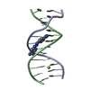
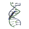
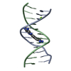
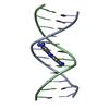
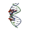
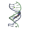
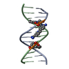
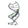
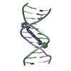
- PDB-360d: STRUCTURE OF 2,5-BIS{[4-(N-ETHYLAMIDINO)PHENYL]}FURAN COMPLEXED T... -
+ Open data

- Basic information
| Entry | Database: PDB / ID: 360d | ||||||||||||||||||||
|---|---|---|---|---|---|---|---|---|---|---|---|---|---|---|---|---|---|---|---|---|---|
| Title | STRUCTURE OF 2,5-BIS{[4-(N-ETHYLAMIDINO)PHENYL]}FURAN COMPLEXED TO 5'-D(CPGPCPGPAPAPTPTPCPGPCPG)-3'. A MINOR GROOVE DRUG COMPLEX, SHOWING PATTERNS OF GROOVE HYDRATION | ||||||||||||||||||||
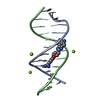
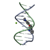
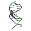
 Components Components | DNA (5'-D(* Keywords Keywords DNA / B-DNA / DOUBLE HELIX / COMPLEXED WITH DRUG DNA / B-DNA / DOUBLE HELIX / COMPLEXED WITH DRUGFunction / homology | Chem-BPF / | DNA / DNA (> 10) Function and homology information Function and homology informationBiological species | synthetic construct (others) | Method | X-RAY DIFFRACTION / MOLECULAR REPLACEMENT / Resolution: 1.85 Å  Authors AuthorsGuerri, A. / Simpson, I.J. / Neidle, S. |  CitationJournal: Nucleic Acids Res. / Year: 1998 CitationJournal: Nucleic Acids Res. / Year: 1998Title: Visualisation of extensive water ribbons and networks in a DNA minor-groove drug complex. Authors: Guerri, A. / Simpson, I.J. / Neidle, S. History |
|
- Structure visualization
| Structure viewer | Molecule: MolmilJmol/JSmol |
|---|
- Downloads & links
-Download
| PDBx/mmCIF format | 360d.cif.gz | 28.2 KB | Display | PDBx/mmCIF format |
|---|---|---|---|---|
| PDB format | pdb360d.ent.gz | 19.2 KB | Display | PDB format |
| PDBx/mmJSON format | 360d.json.gz | Tree view | PDBx/mmJSON format | |
| Others |  Other downloads Other downloads |
-Validation report
| Arichive directory | https://data.pdbj.org/pub/pdb/validation_reports/60/360dftp://data.pdbj.org/pub/pdb/validation_reports/60/360d | HTTPS FTP |
|---|
-Related structure data
| Related structure data | 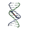 289dS S: Starting model for refinement |
|---|---|
| Similar structure data |


-Links
 PDBj
PDBj


- Assembly
| Deposited unit | 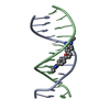
| ||||||||
|---|---|---|---|---|---|---|---|---|---|
| 1 |
| ||||||||
| Unit cell |
|
-Components
| #1: DNA chain | Mass: 3663.392 Da / Num. of mol.: 2 / Source method: obtained synthetically / Source: (synth.) synthetic construct (others) #2: Chemical | ChemComp-MG / |   Mass: 24.305 Da / Num. of mol.: 1 / Source method: obtained synthetically / Formula: Mg Mass: 24.305 Da / Num. of mol.: 1 / Source method: obtained synthetically / Formula: Mg#3: Chemical | ChemComp-BPF / |   Mass: 362.468 Da / Num. of mol.: 1 / Source method: obtained synthetically / Formula: C22H26N4O Mass: 362.468 Da / Num. of mol.: 1 / Source method: obtained synthetically / Formula: C22H26N4O#4: Water | ChemComp-HOH / | Water Mass: 18.015 Da / Num. of mol.: 219 / Source method: isolated from a natural source / Formula: H2O Mass: 18.015 Da / Num. of mol.: 219 / Source method: isolated from a natural source / Formula: H2O |
|---|
-Experimental details
-Experiment
| Experiment | Method: X-RAY DIFFRACTION / Number of used crystals: 1 |
|---|
- Sample preparation
| Crystal | Density Matthews: 2.18 Å3/Da / Density % sol: 43.48 % | ||||||||||||||||||||||||||||||||||||||||||||||||
|---|---|---|---|---|---|---|---|---|---|---|---|---|---|---|---|---|---|---|---|---|---|---|---|---|---|---|---|---|---|---|---|---|---|---|---|---|---|---|---|---|---|---|---|---|---|---|---|---|---|
| Crystal grow | Temperature: 288 K / Method: vapor diffusion / pH: 7 / Details: pH 7.00, VAPOR DIFFUSION, temperature 288.0K | ||||||||||||||||||||||||||||||||||||||||||||||||
| Components of the solutions |
| ||||||||||||||||||||||||||||||||||||||||||||||||
| Crystal grow | *PLUS Temperature: 15 ℃ / pH: 7 / Method: vapor diffusion, hanging drop | ||||||||||||||||||||||||||||||||||||||||||||||||
| Components of the solutions | *PLUS
|
-Data collection
| Diffraction | Mean temperature: 100 K |
|---|---|
| Diffraction source | Source: ROTATING ANODE / Type: RIGAKU |
| Detector | Type: RIGAKU RAXIS II / Detector: IMAGE PLATE / Date: Jul 1, 1997 / Details: MIRROR |
| Radiation | Monochromatic (M) / Laue (L): M / Scattering type: x-ray |
| Radiation wavelength | Relative weight: 1 |
| Reflection | Resolution: 1.85→7 Å / Num. obs: 5634 / % possible obs: 91.44 % / Observed criterion σ(I): 4 / Rmerge(I) obs: 0.022 / Net I/σ(I): 33.18 |
| Reflection shell | Resolution: 1.854→1.9 Å / Mean I/σ(I) obs: 14.67 / % possible all: 80.7 |
| Reflection | *PLUS % possible obs: 96.2 % / Num. measured all: 36613 / Rmerge(I) obs: 0.034 |
- Processing
| Software |
| ||||||||||||||||
|---|---|---|---|---|---|---|---|---|---|---|---|---|---|---|---|---|---|
| Refinement | Method to determine structure: MOLECULAR REPLACEMENT Starting model: NDB ENTRY GDL045 (PDB: 289D) Resolution: 1.85→7 Å / Num. parameters: 2904 / Num. restraintsaints: 3734 / Cross valid method: NONE / σ(F): 4 StereochEM target val spec case: STEREOCHEMISTRY OF LIGAND FROM MM CALCULATIONS Stereochemistry target values: PARKINSON ET AL.
| ||||||||||||||||
| Solvent computation | Solvent model: MOEWS & KRETSINGER, J.MOL.BIOL.91(1973)201-22 | ||||||||||||||||
| Refine analyze | Num. disordered residues: 0 / Occupancy sum hydrogen: 260 / Occupancy sum non hydrogen: 733 | ||||||||||||||||
| Refinement step | Cycle: LAST / Resolution: 1.85→7 Å
| ||||||||||||||||
| Software | *PLUS Name: SHELXL-97 / Classification: refinement | ||||||||||||||||
| Refinement | *PLUS Highest resolution: 1.85 Å / Lowest resolution: 7 Å / σ(F): 4 / Rfactor obs: 0.169 / Rfactor Rfree: 0.227 / Rfactor Rwork: 0.17 | ||||||||||||||||
| Solvent computation | *PLUS | ||||||||||||||||
| Displacement parameters | *PLUS | ||||||||||||||||
| Refine LS restraints | *PLUS
|
