 Movie
Movie Controller

[English] 日本語
 Yorodumi
- PDB-305d: SIDE-BY-SIDE BINDING OF DISTAMYCIN MOLECULES TO D(ICATATIC) IN TH... -
+ Open data

- Basic information
| Entry | Database: PDB / ID: 305d | ||||||||||||||||||
|---|---|---|---|---|---|---|---|---|---|---|---|---|---|---|---|---|---|---|---|
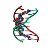
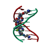
| Title | SIDE-BY-SIDE BINDING OF DISTAMYCIN MOLECULES TO D(ICATATIC) IN THE TETRAGONAL FORM | ||||||||||||||||||
 Components Components | DNA (5'-D(* Keywords Keywords DNA / B-DNA / DOUBLE HELIX / COMPLEXED WITH DRUG / DOUBLE DRUG IN THE MINOR GROOVE DNA / B-DNA / DOUBLE HELIX / COMPLEXED WITH DRUG / DOUBLE DRUG IN THE MINOR GROOVEFunction / homology | DISTAMYCIN A / DNA Function and homology information Function and homology informationMethod | X-RAY DIFFRACTION / DIRECT REFINEMENT / Resolution: 2.17 Å  Authors AuthorsChen, X. / Ramakrishnan, B. / Sundaralingam, M. |  CitationJournal: J.Mol.Biol. / Year: 1997 CitationJournal: J.Mol.Biol. / Year: 1997Title: Crystal structures of the side-by-side binding of distamycin to AT-containing DNA octamers d(ICITACIC) and d(ICATATIC). Authors: Chen, X. / Ramakrishnan, B. / Sundaralingam, M. #1: Journal: Nat.Struct.Biol. / Year: 1994Title: Binding of two Distamycin A Molecules in the Minor Groove of an Alternating B-DNA Authors: Chen, X. / Ramakrishnan, B. / Rao, S.T. / Sundaralingam, M. #2: Journal: Nat.Struct.Biol. / Year: 1995Title: Crystal Structures of B-Form DNA-RNA Chimers Complexed with Distamycin Authors: Chen, X. / Ramakrishnan, B. / Sundaralingam, M. History |
|
- Structure visualization
| Structure viewer | Molecule: MolmilJmol/JSmol |
|---|
- Downloads & links
-Download
| PDBx/mmCIF format | 305d.cif.gz | 16.4 KB | Display | PDBx/mmCIF format |
|---|---|---|---|---|
| PDB format | pdb305d.ent.gz | 9.5 KB | Display | PDB format |
| PDBx/mmJSON format | 305d.json.gz | Tree view | PDBx/mmJSON format | |
| Others |  Other downloads Other downloads |
-Validation report
| Arichive directory | https://data.pdbj.org/pub/pdb/validation_reports/05/305dftp://data.pdbj.org/pub/pdb/validation_reports/05/305d | HTTPS FTP |
|---|
-Related structure data


-Links
 PDBj
PDBj

- Assembly
| Deposited unit | 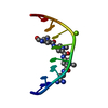
| ||||||||
|---|---|---|---|---|---|---|---|---|---|
| 1 | 
| ||||||||
| Unit cell |
|
-Components
| #1: DNA chain | Mass: 2396.589 Da / Num. of mol.: 1 / Source method: obtained synthetically |
|---|---|
| #2: Chemical | ChemComp-DMY / Distamycin  Mass: 481.508 Da / Num. of mol.: 1 / Source method: obtained synthetically / Formula: C22H27N9O4 / Comment: antibiotic*YM Mass: 481.508 Da / Num. of mol.: 1 / Source method: obtained synthetically / Formula: C22H27N9O4 / Comment: antibiotic*YM |
| #3: Chemical | ChemComp-MG /   Mass: 24.305 Da / Num. of mol.: 1 / Source method: obtained synthetically / Formula: Mg Mass: 24.305 Da / Num. of mol.: 1 / Source method: obtained synthetically / Formula: Mg |
| #4: Water | ChemComp-HOH / Water Mass: 18.015 Da / Num. of mol.: 40 / Source method: isolated from a natural source / Formula: H2O Mass: 18.015 Da / Num. of mol.: 40 / Source method: isolated from a natural source / Formula: H2O |
-Experimental details
-Experiment
| Experiment | Method: X-RAY DIFFRACTION / Number of used crystals: 1 |
|---|
- Sample preparation
| Crystal | Density Matthews: 2.28 Å3/Da / Density % sol: 46.09 % | ||||||||||||||||||||||||||||||||||||||||||||||||
|---|---|---|---|---|---|---|---|---|---|---|---|---|---|---|---|---|---|---|---|---|---|---|---|---|---|---|---|---|---|---|---|---|---|---|---|---|---|---|---|---|---|---|---|---|---|---|---|---|---|
| Crystal grow | Method: vapor diffusion, hanging drop / pH: 7 / Details: pH 7.00, VAPOR DIFFUSION, HANGING DROP / Temp details: ROOM TEMPERATURE | ||||||||||||||||||||||||||||||||||||||||||||||||
| Components of the solutions |
| ||||||||||||||||||||||||||||||||||||||||||||||||
| Crystal grow | *PLUS pH: 7 | ||||||||||||||||||||||||||||||||||||||||||||||||
| Components of the solutions | *PLUS
|
-Data collection
| Diffraction | Mean temperature: 298 K |
|---|---|
| Diffraction source | Source: ROTATING ANODE / Type: RIGAKU |
| Detector | Type: SIEMENS-NICOLET / Detector: AREA DETECTOR / Date: Mar 24, 1994 |
| Radiation | Monochromator: GRAPHITE / Protocol: SINGLE WAVELENGTH / Monochromatic (M) / Laue (L): M / Scattering type: x-ray |
| Radiation wavelength | Relative weight: 1 |
| Reflection | Highest resolution: 2.17 Å / Num. all: 7996 / Num. obs: 1432 / % possible obs: 83 % / Observed criterion σ(I): 0 / Redundancy: 5.6 % / Rsym value: 0.056 |
| Reflection | *PLUS Highest resolution: 2.17 Å / % possible obs: 83 % / Rmerge(I) obs: 0.056 |
- Processing
| Software |
| ||||||||||||||||||||||||||||||||||||||||||||||||||||||||||||
|---|---|---|---|---|---|---|---|---|---|---|---|---|---|---|---|---|---|---|---|---|---|---|---|---|---|---|---|---|---|---|---|---|---|---|---|---|---|---|---|---|---|---|---|---|---|---|---|---|---|---|---|---|---|---|---|---|---|---|---|---|---|
| Refinement | Method to determine structure: DIRECT REFINEMENT Starting model: GDLB51 Resolution: 2.17→8 Å / σ(F): 1
| ||||||||||||||||||||||||||||||||||||||||||||||||||||||||||||
| Displacement parameters | Biso mean: 14.8 Å2 | ||||||||||||||||||||||||||||||||||||||||||||||||||||||||||||
| Refine Biso |
| ||||||||||||||||||||||||||||||||||||||||||||||||||||||||||||
| Refinement step | Cycle: LAST / Resolution: 2.17→8 Å
| ||||||||||||||||||||||||||||||||||||||||||||||||||||||||||||
| Refine LS restraints |
| ||||||||||||||||||||||||||||||||||||||||||||||||||||||||||||
| Software | *PLUS Name: X-PLOR / Classification: refinement | ||||||||||||||||||||||||||||||||||||||||||||||||||||||||||||
| Refinement | *PLUS Highest resolution: 2.17 Å / Lowest resolution: 8 Å / σ(F): 1 | ||||||||||||||||||||||||||||||||||||||||||||||||||||||||||||
| Solvent computation | *PLUS | ||||||||||||||||||||||||||||||||||||||||||||||||||||||||||||
| Displacement parameters | *PLUS Biso mean: 14.8 Å2 | ||||||||||||||||||||||||||||||||||||||||||||||||||||||||||||
| Refine LS restraints | *PLUS
|
