 Movie
Movie Controller

[English] 日本語
 Yorodumi
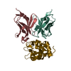
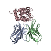
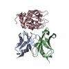
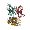
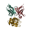
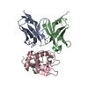
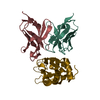
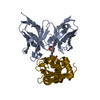
- PDB-2znx: 5-Fluorotryptophan Incorporated ScFv10 Complexed to Hen Egg Lysozyme -
+ Open data

- Basic information
| Entry | Database: PDB / ID: 2znx | ||||||
|---|---|---|---|---|---|---|---|
| Title | 5-Fluorotryptophan Incorporated ScFv10 Complexed to Hen Egg Lysozyme | ||||||
 Components Components |
| ||||||
 Keywords Keywords |  IMMUNE SYSTEM/HYDROLASE / fluorotryptohpan / 5-fluorotryptophan / 19F / Single Chain Fv / Lysozyme / Allergen / Antimicrobial / Bacteriolytic enzyme / Glycosidase / Hydrolase / IMMUNE SYSTEM-HYDROLASE COMPLEX IMMUNE SYSTEM/HYDROLASE / fluorotryptohpan / 5-fluorotryptophan / 19F / Single Chain Fv / Lysozyme / Allergen / Antimicrobial / Bacteriolytic enzyme / Glycosidase / Hydrolase / IMMUNE SYSTEM-HYDROLASE COMPLEX | ||||||
| Function / homology |  Function and homology information Function and homology informationLactose synthesis / Antimicrobial peptides / Neutrophil degranulation / beta-N-acetylglucosaminidase activity / RNA-directed DNA polymerase activity / cell wall macromolecule catabolic process / lysozyme / lysozyme activity / killing of cells of another organism / defense response to Gram-negative bacterium ...Lactose synthesis / Antimicrobial peptides / Neutrophil degranulation / beta-N-acetylglucosaminidase activity / RNA-directed DNA polymerase activity / cell wall macromolecule catabolic process / lysozyme / lysozyme activity / killing of cells of another organism / defense response to Gram-negative bacterium / defense response to Gram-positive bacterium / defense response to bacterium / immune response / endoplasmic reticulum / extracellular space / identical protein binding / cytoplasmSimilarity search - Function | ||||||
| Biological species |  Mus musculus (house mouse) Mus musculus (house mouse)synthetic construct (others)  Gallus gallus (chicken) Gallus gallus (chicken) | ||||||
| Method | X-RAY DIFFRACTION / SYNCHROTRON / MOLECULAR REPLACEMENT / Resolution: 2.3 Å | ||||||
 Authors Authors | DeSantis, M.E. / Acchione, M. / Li, M. / Walter, R.L. / Wlodawer, A. / Smith-Gill, S. | ||||||
 Citation Citation | Journal: Biochemistry / Year: 2012 Title: Specific fluorine labeling of the HyHEL10 antibody affects antigen binding and dynamics Authors: Acchione, M. / Lee, Y.C. / DeSantis, M.E. / Lipschultz, C.A. / Wlodawer, A. / Li, M. / Shanmuganathan, A. / Walter, R.L. / Smith-Gill, S. / Barchi, J.J. | ||||||
| History |
|
- Structure visualization
| Structure viewer | Molecule: MolmilJmol/JSmol |
|---|
- Downloads & links
-Download
| PDBx/mmCIF format | 2znx.cif.gz | 155.5 KB | Display | PDBx/mmCIF format |
|---|---|---|---|---|
| PDB format | pdb2znx.ent.gz | 122.9 KB | Display | PDB format |
| PDBx/mmJSON format | 2znx.json.gz | Tree view | PDBx/mmJSON format | |
| Others |  Other downloads Other downloads |
-Validation report
| Arichive directory | https://data.pdbj.org/pub/pdb/validation_reports/zn/2znxftp://data.pdbj.org/pub/pdb/validation_reports/zn/2znx | HTTPS FTP |
|---|
-Related structure data
| Related structure data |  2znwC  2dqjS C: citing same article ( S: Starting model for refinement |
|---|---|
| Similar structure data |


-Links
 PDBj
PDBj


- Assembly
| Deposited unit | 
| |||||||||||||||||||||||||||||||||||||||||||||||||||||||||||||||||||||||||||||||||||||||||||||||
|---|---|---|---|---|---|---|---|---|---|---|---|---|---|---|---|---|---|---|---|---|---|---|---|---|---|---|---|---|---|---|---|---|---|---|---|---|---|---|---|---|---|---|---|---|---|---|---|---|---|---|---|---|---|---|---|---|---|---|---|---|---|---|---|---|---|---|---|---|---|---|---|---|---|---|---|---|---|---|---|---|---|---|---|---|---|---|---|---|---|---|---|---|---|---|---|---|
| 1 | 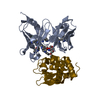
| |||||||||||||||||||||||||||||||||||||||||||||||||||||||||||||||||||||||||||||||||||||||||||||||
| 2 | 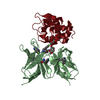
| |||||||||||||||||||||||||||||||||||||||||||||||||||||||||||||||||||||||||||||||||||||||||||||||
| Unit cell |
| |||||||||||||||||||||||||||||||||||||||||||||||||||||||||||||||||||||||||||||||||||||||||||||||
| Noncrystallographic symmetry (NCS) | NCS domain:
NCS domain segments: Component-ID: 1 / Refine code: 4
NCS ensembles :
|
-Components
| #1: Antibody | Single-chain variable fragment Mass: 26270.408 Da / Num. of mol.: 2 Source method: isolated from a genetically manipulated source Details: This molecule contains antibody light chain(residues 1-107), linker(residues 108-122) and antibody heavy chain(residues 123-236) Source: (gene. exp.) Mus musculus (house mouse), (gene. exp.) synthetic construct (others)Plasmid: pET30 / Production host:  Escherichia coli (E. coli) / Strain (production host): CT19 BL21(DE3) / References: UniProt: Q65ZI1*PLUS Escherichia coli (E. coli) / Strain (production host): CT19 BL21(DE3) / References: UniProt: Q65ZI1*PLUS#2: Protein | Mass: 14331.160 Da / Num. of mol.: 2 / Source method: isolated from a natural source / Source: (natural) Gallus gallus (chicken) / Tissue: egg white / References: UniProt: P00698, lysozyme#3: Chemical | Polyethylene glycol  Mass: 252.305 Da / Num. of mol.: 2 / Source method: obtained synthetically / Formula: C11H24O6 Mass: 252.305 Da / Num. of mol.: 2 / Source method: obtained synthetically / Formula: C11H24O6#4: Water | ChemComp-HOH / | Water Mass: 18.015 Da / Num. of mol.: 311 / Source method: isolated from a natural source / Formula: H2O Mass: 18.015 Da / Num. of mol.: 311 / Source method: isolated from a natural source / Formula: H2O |
|---|
-Experimental details
-Experiment
| Experiment | Method: X-RAY DIFFRACTION / Number of used crystals: 1 |
|---|
- Sample preparation
| Crystal | Density Matthews: 2.78 Å3/Da / Density % sol: 55.79 % |
|---|---|
| Crystal grow | Temperature: 293 K / Method: vapor diffusion, sitting drop / pH: 8.5 Details: 30% PEG400, 0.2M Na Citrate; 0.1M Tris, pH 8.5, VAPOR DIFFUSION, SITTING DROP, temperature 293K |
-Data collection
| Diffraction | Mean temperature: 100 K |
|---|---|
| Diffraction source | Source: SYNCHROTRON / Site: APS  / Beamline: 22-ID / Wavelength: 1 Å / Beamline: 22-ID / Wavelength: 1 Å |
| Detector | Type: MARMOSAIC 300 mm CCD / Detector: CCD / Date: Dec 18, 2007 / Details: mirrors |
| Radiation | Monochromator: graphite / Protocol: SINGLE WAVELENGTH / Monochromatic (M) / Laue (L): M / Scattering type: x-ray |
| Radiation wavelength | Wavelength: 1 Å / Relative weight: 1 |
| Reflection | Resolution: 2.2→50 Å / Num. all: 47378 / Num. obs: 47378 / % possible obs: 99.9 % / Observed criterion σ(I): -3 / Redundancy: 9 % / Rsym value: 0.12 / Net I/σ(I): 22.5 |
| Reflection shell | Resolution: 2.2→2.28 Å / Redundancy: 8.8 % / Mean I/σ(I) obs: 4.2 / Num. unique all: 4648 / Rsym value: 0.786 / % possible all: 100 |
- Processing
| Software |
| |||||||||||||||||||||||||||||||||||||||||||||||||||||||||||||||||
|---|---|---|---|---|---|---|---|---|---|---|---|---|---|---|---|---|---|---|---|---|---|---|---|---|---|---|---|---|---|---|---|---|---|---|---|---|---|---|---|---|---|---|---|---|---|---|---|---|---|---|---|---|---|---|---|---|---|---|---|---|---|---|---|---|---|---|
| Refinement | Method to determine structure: MOLECULAR REPLACEMENT Starting model: PDB entry 2DQJ Resolution: 2.3→50 Å / Cor.coef. Fo:Fc: 0.94 / Cor.coef. Fo:Fc free: 0.908 / SU B: 5.36 / SU ML: 0.135 / Cross valid method: THROUGHOUT / σ(F): 0 / ESU R: 0.272 / ESU R Free: 0.209 / Stereochemistry target values: MAXIMUM LIKELIHOOD / Details: HYDROGENS HAVE BEEN ADDED IN THE RIDING POSITIONS
| |||||||||||||||||||||||||||||||||||||||||||||||||||||||||||||||||
| Solvent computation | Ion probe radii: 0.8 Å / Shrinkage radii: 0.8 Å / VDW probe radii: 1.2 Å / Solvent model: MASK | |||||||||||||||||||||||||||||||||||||||||||||||||||||||||||||||||
| Displacement parameters | Biso mean: 26.604 Å2
| |||||||||||||||||||||||||||||||||||||||||||||||||||||||||||||||||
| Refinement step | Cycle: LAST / Resolution: 2.3→50 Å
| |||||||||||||||||||||||||||||||||||||||||||||||||||||||||||||||||
| Refine LS restraints |
| |||||||||||||||||||||||||||||||||||||||||||||||||||||||||||||||||
| Refine LS restraints NCS | Dom-ID: 1 / Refine-ID: X-RAY DIFFRACTION
| |||||||||||||||||||||||||||||||||||||||||||||||||||||||||||||||||
| LS refinement shell | Resolution: 2.3→2.36 Å / Total num. of bins used: 20
|
