 Movie
Movie Controller


+ Open data

- Basic information
| Entry | Database: PDB / ID: 2zhv | ||||||
|---|---|---|---|---|---|---|---|
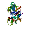
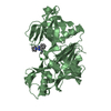
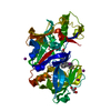
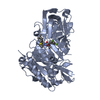
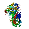
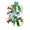
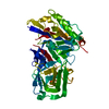
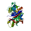
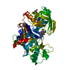
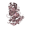
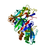
| Title | Crystal structure of BACE1 at pH 7.0 | ||||||
 Components Components | Beta-secretase 1 | ||||||
 Keywords Keywords | HYDROLASE / pH 7.0 / Alternative splicing / Aspartyl protease / Glycoprotein / Membrane / Protease / Transmembrane / Zymogen | ||||||
| Function / homology |  Function and homology informationmemapsin 2 / Golgi-associated vesicle lumen / signaling receptor ligand precursor processing / beta-aspartyl-peptidase activity / amyloid precursor protein catabolic process / amyloid-beta formation / membrane protein ectodomain proteolysis / cellular response to manganese ion / amyloid-beta metabolic process / prepulse inhibition ...memapsin 2 / Golgi-associated vesicle lumen / signaling receptor ligand precursor processing / beta-aspartyl-peptidase activity / amyloid precursor protein catabolic process / amyloid-beta formation / membrane protein ectodomain proteolysis / cellular response to manganese ion / amyloid-beta metabolic process / prepulse inhibition / detection of mechanical stimulus involved in sensory perception of pain / cellular response to copper ion / presynaptic modulation of chemical synaptic transmission / hippocampal mossy fiber to CA3 synapse / multivesicular body / response to lead ion / trans-Golgi network / recycling endosome / protein processing / cellular response to amyloid-beta / positive regulation of neuron apoptotic process / synaptic vesicle / late endosome / peptidase activity / amyloid-beta binding / endopeptidase activity / amyloid fibril formation / lysosome / aspartic-type endopeptidase activity / early endosome / endosome membrane / endosome / membrane raft / Amyloid fiber formation / endoplasmic reticulum lumen / axon / neuronal cell body / dendrite / Golgi apparatus / enzyme binding / cell surface / proteolysis / membrane / plasma membrane Function and homology informationmemapsin 2 / Golgi-associated vesicle lumen / signaling receptor ligand precursor processing / beta-aspartyl-peptidase activity / amyloid precursor protein catabolic process / amyloid-beta formation / membrane protein ectodomain proteolysis / cellular response to manganese ion / amyloid-beta metabolic process / prepulse inhibition ...memapsin 2 / Golgi-associated vesicle lumen / signaling receptor ligand precursor processing / beta-aspartyl-peptidase activity / amyloid precursor protein catabolic process / amyloid-beta formation / membrane protein ectodomain proteolysis / cellular response to manganese ion / amyloid-beta metabolic process / prepulse inhibition / detection of mechanical stimulus involved in sensory perception of pain / cellular response to copper ion / presynaptic modulation of chemical synaptic transmission / hippocampal mossy fiber to CA3 synapse / multivesicular body / response to lead ion / trans-Golgi network / recycling endosome / protein processing / cellular response to amyloid-beta / positive regulation of neuron apoptotic process / synaptic vesicle / late endosome / peptidase activity / amyloid-beta binding / endopeptidase activity / amyloid fibril formation / lysosome / aspartic-type endopeptidase activity / early endosome / endosome membrane / endosome / membrane raft / Amyloid fiber formation / endoplasmic reticulum lumen / axon / neuronal cell body / dendrite / Golgi apparatus / enzyme binding / cell surface / proteolysis / membrane / plasma membraneSimilarity search - Function | ||||||
| Biological species |  Homo sapiens (human) Homo sapiens (human) | ||||||
| Method | X-RAY DIFFRACTION / SYNCHROTRON / MOLECULAR REPLACEMENT / Resolution: 1.85 Å | ||||||
 Authors Authors | Shimizu, H. / Nukina, N. | ||||||
 Citation Citation | Journal: Mol.Cell.Biol. / Year: 2008 Title: Crystal structure of an active form of BACE1, an enzyme responsible for amyloid beta protein production Authors: Shimizu, H. / Tosaki, A. / Kaneko, K. / Hisano, T. / Sakurai, T. / Nukina, N. | ||||||
| History |
|
- Structure visualization
| Structure viewer | Molecule: MolmilJmol/JSmol |
|---|
- Downloads & links
-Download
| PDBx/mmCIF format | 2zhv.cif.gz | 93.1 KB | Display | PDBx/mmCIF format |
|---|---|---|---|---|
| PDB format | pdb2zhv.ent.gz | 69.5 KB | Display | PDB format |
| PDBx/mmJSON format | 2zhv.json.gz | Tree view | PDBx/mmJSON format | |
| Others |  Other downloads Other downloads |
-Validation report
| Arichive directory | https://data.pdbj.org/pub/pdb/validation_reports/zh/2zhvftp://data.pdbj.org/pub/pdb/validation_reports/zh/2zhv | HTTPS FTP |
|---|
-Related structure data
| Related structure data |  2zhrC 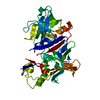 2zhsC 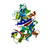 2zhtC  2zhuC 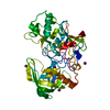 1w50S C: citing same article ( S: Starting model for refinement |
|---|---|
| Similar structure data |


-Links
 PDBj
PDBj

- Assembly
| Deposited unit | 
| ||||||||
|---|---|---|---|---|---|---|---|---|---|
| 1 |
| ||||||||
| Unit cell |
|
-Components
| #1: Protein | / Beta-site APP cleaving enzyme 1 / Beta-site amyloid precursor protein cleaving enzyme 1 / Membrane- ...Beta-site APP cleaving enzyme 1 / Beta-site amyloid precursor protein cleaving enzyme 1 / Membrane-associated aspartic protease 2 / Memapsin-2 / Aspartyl protease 2 / Asp 2 / ASP2 / BACE1 Mass: 45900.500 Da / Num. of mol.: 1 / Fragment: catalytic domain, UNP residues 45-454 Source method: isolated from a genetically manipulated source Source: (gene. exp.) Homo sapiens (human) / Gene: BACE1 / Plasmid: pet11a / Species (production host): Escherichia coli / Production host:  Escherichia coli BL21 (bacteria) / Strain (production host): BL21 / References: UniProt: P56817, memapsin 2 Escherichia coli BL21 (bacteria) / Strain (production host): BL21 / References: UniProt: P56817, memapsin 2 |
|---|---|
| #2: Water | ChemComp-HOH / Water Mass: 18.015 Da / Num. of mol.: 265 / Source method: isolated from a natural source / Formula: H2O Mass: 18.015 Da / Num. of mol.: 265 / Source method: isolated from a natural source / Formula: H2O |
-Experimental details
-Experiment
| Experiment | Method: X-RAY DIFFRACTION / Number of used crystals: 1 |
|---|
- Sample preparation
| Crystal | Density Matthews: 2.81 Å3/Da / Density % sol: 56.19 % |
|---|---|
| Crystal grow | Temperature: 293 K / Method: vapor diffusion, sitting drop / pH: 6.5 Details: 22% PEG 5000 MME, 0.2M Sodium Acetate pH 6.5, 0.2M Ammonium Iodide, VAPOR DIFFUSION, SITTING DROP, temperature 293K |
-Data collection
| Diffraction | Mean temperature: 100 K |
|---|---|
| Diffraction source | Source: SYNCHROTRON / Site: SPring-8  / Beamline: BL44B2 / Wavelength: 1 Å / Beamline: BL44B2 / Wavelength: 1 Å |
| Detector | Type: ADSC QUANTUM 210 / Detector: CCD / Date: Jun 28, 2006 |
| Radiation | Protocol: SINGLE WAVELENGTH / Monochromatic (M) / Laue (L): M / Scattering type: x-ray |
| Radiation wavelength | Wavelength: 1 Å / Relative weight: 1 |
| Reflection | Resolution: 1.85→100 Å / Num. obs: 44019 / % possible obs: 95.7 % / Redundancy: 5 % / Rsym value: 0.048 / Net I/σ(I): 40.1 |
| Reflection shell | Resolution: 1.85→1.92 Å / Redundancy: 2 % / Mean I/σ(I) obs: 3.1 / Num. unique all: 3739 / Rsym value: 0.341 / % possible all: 83.3 |
- Processing
| Software |
| ||||||||||||||||||||
|---|---|---|---|---|---|---|---|---|---|---|---|---|---|---|---|---|---|---|---|---|---|
| Refinement | Method to determine structure: MOLECULAR REPLACEMENT Starting model: PDB ENTRY 1W50 Resolution: 1.85→49.01 Å / Rfactor Rfree error: 0.006 / Data cutoff high absF: 3013944.27 / Data cutoff low absF: 0 / Isotropic thermal model: RESTRAINED / Cross valid method: THROUGHOUT / σ(F): 0
| ||||||||||||||||||||
| Solvent computation | Solvent model: FLAT MODEL / Bsol: 61.0145 Å2 / ksol: 0.361145 e/Å3 | ||||||||||||||||||||
| Displacement parameters | Biso mean: 35.2 Å2
| ||||||||||||||||||||
| Refinement step | Cycle: LAST / Resolution: 1.85→49.01 Å
| ||||||||||||||||||||
| Refine LS restraints |
| ||||||||||||||||||||
| LS refinement shell | Highest resolution: 1.85 Å / Total num. of bins used: 6 /
| ||||||||||||||||||||
| Xplor file |
|
