 Movie
Movie Controller

[English] 日本語
 Yorodumi
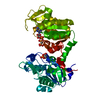
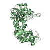
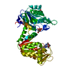
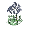
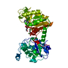
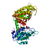
- PDB-2zgv: Crystal Structure of human phosphoglycerate kinase bound to D-ADP -
+ Open data

- Basic information
| Entry | Database: PDB / ID: 2zgv | ||||||
|---|---|---|---|---|---|---|---|
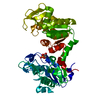
| Title | Crystal Structure of human phosphoglycerate kinase bound to D-ADP | ||||||
 Components Components | Phosphoglycerate kinase 1 | ||||||
 Keywords Keywords | TRANSFERASE / PROTEIN-NUCLEOTIDE COMPLEX / Acetylation / ATP-binding / Cytoplasm / Disease mutation / Glycolysis / Hereditary hemolytic anemia / Kinase / Nucleotide-binding / Phosphoprotein / Polymorphism | ||||||
| Function / homology |  Function and homology information Function and homology informationManipulation of host energy metabolism / phosphoglycerate kinase / phosphoglycerate kinase activity / protein-disulfide reductase (NAD(P)H) activity / Gluconeogenesis / canonical glycolysis / Glycolysis / plasminogen activation / epithelial cell differentiation / negative regulation of angiogenesis ...Manipulation of host energy metabolism / phosphoglycerate kinase / phosphoglycerate kinase activity / protein-disulfide reductase (NAD(P)H) activity / Gluconeogenesis / canonical glycolysis / Glycolysis / plasminogen activation / epithelial cell differentiation / negative regulation of angiogenesis / gluconeogenesis / glycolytic process / ADP binding / cellular response to hypoxia / membrane raft / phosphorylation / extracellular space / extracellular exosome / ATP binding / membrane / cytosolSimilarity search - Function | ||||||
| Biological species |  Homo sapiens (human) Homo sapiens (human) | ||||||
| Method | X-RAY DIFFRACTION / SYNCHROTRON / MOLECULAR REPLACEMENT / Resolution: 2 Å | ||||||
 Authors Authors | Arold, S.T. / Gondeau, C. / Lionne, C. / Chaloin, L. | ||||||
 Citation Citation | Journal: Nucleic Acids Res. / Year: 2008 Title: Molecular basis for the lack of enantioselectivity of human 3-phosphoglycerate kinase Authors: Gondeau, C. / Chaloin, L. / Lallemand, P. / Roy, B. / Perigaud, C. / Barman, T. / Varga, A. / Vas, M. / Lionne, C. / Arold, S.T. | ||||||
| History |
|
- Structure visualization
| Structure viewer | Molecule: MolmilJmol/JSmol |
|---|
- Downloads & links
-Download
| PDBx/mmCIF format | 2zgv.cif.gz | 95 KB | Display | PDBx/mmCIF format |
|---|---|---|---|---|
| PDB format | pdb2zgv.ent.gz | 69.5 KB | Display | PDB format |
| PDBx/mmJSON format | 2zgv.json.gz | Tree view | PDBx/mmJSON format | |
| Others |  Other downloads Other downloads |
-Validation report
| Arichive directory | https://data.pdbj.org/pub/pdb/validation_reports/zg/2zgvftp://data.pdbj.org/pub/pdb/validation_reports/zg/2zgv | HTTPS FTP |
|---|
-Related structure data
| Related structure data | 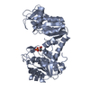 3c39C  3c3aC 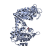 3c3bC 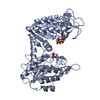 3c3cC  1vjcS C: citing same article ( S: Starting model for refinement |
|---|---|
| Similar structure data |


-Links
 PDBj
PDBj

- Assembly
| Deposited unit | 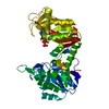
| ||||||||
|---|---|---|---|---|---|---|---|---|---|
| 1 |
| ||||||||
| Unit cell |
|
-Components
| #1: Protein | / 3-Phosphoglycerate Kinase / Primer recognition protein 2 / PRP 2 / Cell migration-inducing gene 10 protein Mass: 44954.895 Da / Num. of mol.: 1 Source method: isolated from a genetically manipulated source Source: (gene. exp.) Homo sapiens (human) / Gene: PGK1 / Plasmid: pET28a / Production host:  Escherichia coli (E. coli) / Strain (production host): BL21 (DE3)/pDIA17 / References: UniProt: P00558, phosphoglycerate kinase Escherichia coli (E. coli) / Strain (production host): BL21 (DE3)/pDIA17 / References: UniProt: P00558, phosphoglycerate kinase |
|---|---|
| #2: Chemical | ChemComp-ADP / Adenosine diphosphate  Mass: 427.201 Da / Num. of mol.: 1 / Source method: obtained synthetically / Formula: C10H15N5O10P2 / Comment: ADP, energy-carrying molecule*YM Mass: 427.201 Da / Num. of mol.: 1 / Source method: obtained synthetically / Formula: C10H15N5O10P2 / Comment: ADP, energy-carrying molecule*YM |
| #3: Water | ChemComp-HOH / Water Mass: 18.015 Da / Num. of mol.: 234 / Source method: isolated from a natural source / Formula: H2O Mass: 18.015 Da / Num. of mol.: 234 / Source method: isolated from a natural source / Formula: H2O |
-Experimental details
-Experiment
| Experiment | Method: X-RAY DIFFRACTION / Number of used crystals: 1 |
|---|
- Sample preparation
| Crystal | Density Matthews: 2.14 Å3/Da / Density % sol: 42.6 % |
|---|---|
| Crystal grow | Temperature: 291 K / Method: vapor diffusion, hanging drop / pH: 8.3 Details: 2.6M NaKPO4, pH 8.3, VAPOR DIFFUSION, HANGING DROP, temperature 291K |
-Data collection
| Diffraction | Mean temperature: 100 K |
|---|---|
| Diffraction source | Source: SYNCHROTRON / Site: ESRF  / Beamline: ID14-4 / Wavelength: 0.934 Å / Beamline: ID14-4 / Wavelength: 0.934 Å |
| Detector | Detector: AREA DETECTOR / Date: May 22, 2007 |
| Radiation | Protocol: SINGLE WAVELENGTH / Monochromatic (M) / Laue (L): M / Scattering type: x-ray |
| Radiation wavelength | Wavelength: 0.934 Å / Relative weight: 1 |
| Reflection | Resolution: 2→45.22 Å / Num. all: 16987 / Num. obs: 16987 / % possible obs: 0.69 % / Observed criterion σ(F): 0 / Observed criterion σ(I): 0 / Redundancy: 2.6 % / Biso Wilson estimate: 19.2 Å2 / Rmerge(I) obs: 0.048 / Net I/σ(I): 11.3 |
| Reflection shell | Resolution: 2→2.11 Å / Redundancy: 2 % / Mean I/σ(I) obs: 2.9 / Num. unique all: 1136 / Rsym value: 0.323 / % possible all: 0.31 |
- Processing
| Software |
| |||||||||||||||||||||||||||||||||||||||||||||||||||||||||||||||||
|---|---|---|---|---|---|---|---|---|---|---|---|---|---|---|---|---|---|---|---|---|---|---|---|---|---|---|---|---|---|---|---|---|---|---|---|---|---|---|---|---|---|---|---|---|---|---|---|---|---|---|---|---|---|---|---|---|---|---|---|---|---|---|---|---|---|---|
| Refinement | Method to determine structure: MOLECULAR REPLACEMENT Starting model: 1VJC Resolution: 2→45.22 Å / Cor.coef. Fo:Fc: 0.947 / Cor.coef. Fo:Fc free: 0.887 / SU B: 5.383 / SU ML: 0.15 / Cross valid method: THROUGHOUT / ESU R: 0.371 / ESU R Free: 0.26 / Stereochemistry target values: MAXIMUM LIKELIHOOD / Details: HYDROGENS HAVE BEEN ADDED IN THE RIDING POSITIONS
| |||||||||||||||||||||||||||||||||||||||||||||||||||||||||||||||||
| Solvent computation | Ion probe radii: 0.8 Å / Shrinkage radii: 0.8 Å / VDW probe radii: 1.4 Å / Solvent model: MASK | |||||||||||||||||||||||||||||||||||||||||||||||||||||||||||||||||
| Displacement parameters | Biso mean: 19.739 Å2
| |||||||||||||||||||||||||||||||||||||||||||||||||||||||||||||||||
| Refinement step | Cycle: LAST / Resolution: 2→45.22 Å
| |||||||||||||||||||||||||||||||||||||||||||||||||||||||||||||||||
| Refine LS restraints |
| |||||||||||||||||||||||||||||||||||||||||||||||||||||||||||||||||
| LS refinement shell | Resolution: 2→2.052 Å / Total num. of bins used: 20
|
