 Movie
Movie Controller

[English] 日本語
 Yorodumi
- PDB-2p9t: Crystal Structure of Phosphoglycerate Kinase-2 bound to 3-phospho... -
+ Open data

- Basic information
| Entry | Database: PDB / ID: 2p9t | ||||||
|---|---|---|---|---|---|---|---|
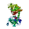
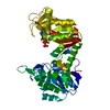
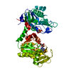
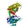
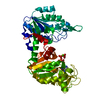
| Title | Crystal Structure of Phosphoglycerate Kinase-2 bound to 3-phosphoglycerate | ||||||
 Components Components | Phosphoglycerate kinase, testis specific | ||||||
 Keywords Keywords | TRANSFERASE / phosphoglycerate kinse / enzyme-substrate complex | ||||||
| Function / homology |  Function and homology information Function and homology informationsperm fibrous sheath / Glycolysis / Gluconeogenesis / phosphoglycerate kinase / phosphoglycerate kinase activity / flagellated sperm motility / gluconeogenesis / glycolytic process / ADP binding / cilium ...sperm fibrous sheath / Glycolysis / Gluconeogenesis / phosphoglycerate kinase / phosphoglycerate kinase activity / flagellated sperm motility / gluconeogenesis / glycolytic process / ADP binding / cilium / phosphorylation / extracellular exosome / ATP binding / cytosolSimilarity search - Function | ||||||
| Biological species |  Mus musculus (house mouse) Mus musculus (house mouse) | ||||||
| Method | X-RAY DIFFRACTION / MOLECULAR REPLACEMENT / Resolution: 2 Å | ||||||
 Authors Authors | Sawyer, G.M. / Monzingo, A.F. / Poteet, E.C. / Robertus, J.D. | ||||||
 Citation Citation | Journal: Proteins / Year: 2007 Title: X-ray analysis of phosphoglycerate kinase 2, a sperm-specific isoform from Mus musculus. Authors: Sawyer, G.M. / Monzingo, A.F. / Poteet, E.C. / O'Brien, D.A. / Robertus, J.D. | ||||||
| History |
| ||||||
| Remark 999 | sequence There is a Gln -> Arg sequence conflict at residue 151 in the Uniprot database. Authors ...sequence There is a Gln -> Arg sequence conflict at residue 151 in the Uniprot database. Authors state that Protein sequence used in this PDB entry is from C3H/He strain which has Arg at residue 150, whereas UniProt sequence is from BALB/c strain that has Gln at residue 151. |
- Structure visualization
| Structure viewer | Molecule: MolmilJmol/JSmol |
|---|
- Downloads & links
-Download
| PDBx/mmCIF format | 2p9t.cif.gz | 96.8 KB | Display | PDBx/mmCIF format |
|---|---|---|---|---|
| PDB format | pdb2p9t.ent.gz | 71.9 KB | Display | PDB format |
| PDBx/mmJSON format | 2p9t.json.gz | Tree view | PDBx/mmJSON format | |
| Others |  Other downloads Other downloads |
-Validation report
| Arichive directory | https://data.pdbj.org/pub/pdb/validation_reports/p9/2p9tftp://data.pdbj.org/pub/pdb/validation_reports/p9/2p9t | HTTPS FTP |
|---|
-Related structure data
| Related structure data | 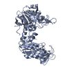 2p9qC 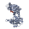 2paaC 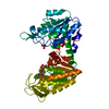 1vjdS C: citing same article ( S: Starting model for refinement |
|---|---|
| Similar structure data |


-Links
 PDBj
PDBj
- Assembly
| Deposited unit | 
| ||||||||
|---|---|---|---|---|---|---|---|---|---|
| 1 |
| ||||||||
| Unit cell |
|
-Components
| #1: Protein | Mass: 44837.836 Da / Num. of mol.: 1 Source method: isolated from a genetically manipulated source Source: (gene. exp.) Mus musculus (house mouse) / Strain: C3H/He / Gene: Pgk2, Pgk-2 / Plasmid: PGK-2-pGEX4T3 / Production host:  Escherichia coli (E. coli) / References: UniProt: P09041, phosphoglycerate kinase Escherichia coli (E. coli) / References: UniProt: P09041, phosphoglycerate kinase |
|---|---|
| #2: Chemical | ChemComp-3PG / 3-Phosphoglyceric acid  Mass: 186.057 Da / Num. of mol.: 1 / Source method: obtained synthetically / Formula: C3H7O7P Mass: 186.057 Da / Num. of mol.: 1 / Source method: obtained synthetically / Formula: C3H7O7P |
| #3: Water | ChemComp-HOH / Water Mass: 18.015 Da / Num. of mol.: 289 / Source method: isolated from a natural source / Formula: H2O Mass: 18.015 Da / Num. of mol.: 289 / Source method: isolated from a natural source / Formula: H2O |
-Experimental details
-Experiment
| Experiment | Method: X-RAY DIFFRACTION / Number of used crystals: 1 |
|---|
- Sample preparation
| Crystal | Density Matthews: 2.16 Å3/Da / Density % sol: 43.02 % |
|---|---|
| Crystal grow | Temperature: 298 K / Method: vapor diffusion, hanging drop / pH: 8 Details: 10 mM D-3-phosphoglycerate, 30% PEG6000, 0.1 M Tris-Hcl, VAPOR DIFFUSION, HANGING DROP, temperature 298K, pH 8.00 |
-Data collection
| Diffraction | Mean temperature: 100 K |
|---|---|
| Diffraction source | Source: ROTATING ANODE / Type: RIGAKU RUH3R / Wavelength: 1.5418 |
| Detector | Type: RIGAKU RAXIS IV / Detector: IMAGE PLATE / Date: May 5, 2006 / Details: BLUE MAX-FLUX CONFOCAL OPTICAL SYSTEM |
| Radiation | Monochromator: BLUE MAX-FLUX CONFOCAL OPTICAL SYSTEM / Protocol: SINGLE WAVELENGTH / Monochromatic (M) / Laue (L): M / Scattering type: x-ray |
| Radiation wavelength | Wavelength: 1.5418 Å / Relative weight: 1 |
| Reflection | Resolution: 2→50 Å / Num. obs: 24578 / % possible obs: 99.3 % / Redundancy: 6.4 % / Rmerge(I) obs: 0.062 / Net I/σ(I): 14.6 |
| Reflection shell | Resolution: 2→2.1 Å / Redundancy: 6 % / Rmerge(I) obs: 0.209 / % possible all: 96.7 |
- Processing
| Software |
| ||||||||||||||||||||||||||||||||||||||||||||||||||||||||||||||||||||||||||||||||
|---|---|---|---|---|---|---|---|---|---|---|---|---|---|---|---|---|---|---|---|---|---|---|---|---|---|---|---|---|---|---|---|---|---|---|---|---|---|---|---|---|---|---|---|---|---|---|---|---|---|---|---|---|---|---|---|---|---|---|---|---|---|---|---|---|---|---|---|---|---|---|---|---|---|---|---|---|---|---|---|---|---|
| Refinement | Method to determine structure: MOLECULAR REPLACEMENT Starting model: PDB ENTRY 1VJD Resolution: 2→50 Å / Cross valid method: THROUGHOUT / σ(F): 0 / Stereochemistry target values: ENGH & HUBER
| ||||||||||||||||||||||||||||||||||||||||||||||||||||||||||||||||||||||||||||||||
| Solvent computation | Bsol: 43.21 Å2 | ||||||||||||||||||||||||||||||||||||||||||||||||||||||||||||||||||||||||||||||||
| Displacement parameters | Biso mean: 24.62 Å2
| ||||||||||||||||||||||||||||||||||||||||||||||||||||||||||||||||||||||||||||||||
| Refinement step | Cycle: LAST / Resolution: 2→50 Å
| ||||||||||||||||||||||||||||||||||||||||||||||||||||||||||||||||||||||||||||||||
| Refine LS restraints |
| ||||||||||||||||||||||||||||||||||||||||||||||||||||||||||||||||||||||||||||||||
| LS refinement shell | Resolution: 2→2.03 Å / Total num. of bins used: 24 | ||||||||||||||||||||||||||||||||||||||||||||||||||||||||||||||||||||||||||||||||
| Xplor file |
|
