 Movie
Movie Controller


+ Open data

- Basic information
| Entry | Database: PDB / ID: 2zfu | ||||||
|---|---|---|---|---|---|---|---|
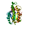
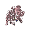
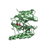
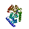
| Title | Structure of the methyltransferase-like domain of nucleomethylin | ||||||
 Components Components | Cerebral protein 1 | ||||||
 Keywords Keywords |  NUCLEAR PROTEIN / NUCLEOLAR PROTEIN / SAM-BINDING PROTEIN / protein structure / Nucleus / Phosphoprotein NUCLEAR PROTEIN / NUCLEOLAR PROTEIN / SAM-BINDING PROTEIN / protein structure / Nucleus / Phosphoprotein | ||||||
| Function / homology |  Function and homology information Function and homology informationregulation of transcription by glucose / regulation of cell cycle => GO:0051726 / rDNA heterochromatin / rDNA heterochromatin formation / chromatin silencing complex / S-adenosylmethionine-dependent methyltransferase activity / intrinsic apoptotic signaling pathway by p53 class mediator / cellular response to glucose starvation / methylated histone binding / Transferases; Transferring one-carbon groups; Methyltransferases ...regulation of transcription by glucose / regulation of cell cycle => GO:0051726 / rDNA heterochromatin / rDNA heterochromatin formation / chromatin silencing complex / S-adenosylmethionine-dependent methyltransferase activity / intrinsic apoptotic signaling pathway by p53 class mediator / cellular response to glucose starvation / methylated histone binding / Transferases; Transferring one-carbon groups; Methyltransferases / SIRT1 negatively regulates rRNA expression / rRNA processing / methylation / nucleolus / RNA binding / nucleoplasm / plasma membrane / cytosolSimilarity search - Function | ||||||
| Biological species |  Homo sapiens (human) Homo sapiens (human) | ||||||
| Method | X-RAY DIFFRACTION / SYNCHROTRON / MAD / Resolution: 2 Å | ||||||
 Authors Authors | Minami, H. / Hashimoto, H. / Murayama, A. / Yanagisawa, J. / Sato, M. / Shimizu, T. | ||||||
 Citation Citation | Journal: Cell(Cambridge,Mass.) / Year: 2008 Title: Epigenetic control of rDNA loci in response to intracellular energy status Authors: Murayama, A. / Ohmori, K. / Fujimura, A. / Minami, H. / Yasuzawa-Tanaka, K. / Kuroda, T. / Oie, S. / Daitoku, H. / Okuwaki, M. / Nagata, K. / Fukamizu, A. / Kimura, K. / Shimizu, T. / Yanagisawa, J. | ||||||
| History |
|
- Structure visualization
| Structure viewer | Molecule: MolmilJmol/JSmol |
|---|
- Downloads & links
-Download
| PDBx/mmCIF format | 2zfu.cif.gz | 101 KB | Display | PDBx/mmCIF format |
|---|---|---|---|---|
| PDB format | pdb2zfu.ent.gz | 76.6 KB | Display | PDB format |
| PDBx/mmJSON format | 2zfu.json.gz | Tree view | PDBx/mmJSON format | |
| Others |  Other downloads Other downloads |
-Validation report
| Arichive directory | https://data.pdbj.org/pub/pdb/validation_reports/zf/2zfuftp://data.pdbj.org/pub/pdb/validation_reports/zf/2zfu | HTTPS FTP |
|---|
-Related structure data
| Similar structure data |
|---|


-Links
 PDBj
PDBj


- Assembly
| Deposited unit | 
| ||||||||
|---|---|---|---|---|---|---|---|---|---|
| 1 | 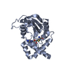
| ||||||||
| 2 | 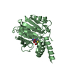
| ||||||||
| Unit cell |
|
-Components
| #1: Protein | Mass: 24297.004 Da / Num. of mol.: 2 Fragment: methyltransferase-like domain, UNP residues 242-456 Source method: isolated from a genetically manipulated source Source: (gene. exp.) Homo sapiens (human) / Gene: KIAA0409 / Plasmid: pGEX4T-2 / Production host:  Escherichia coli (E. coli) / Strain (production host): BL21(DE3) / References: UniProt: O43159 Escherichia coli (E. coli) / Strain (production host): BL21(DE3) / References: UniProt: O43159#2: Chemical | S-Adenosyl-L-homocysteine  Type: L-peptide linking / Mass: 384.411 Da / Num. of mol.: 2 / Source method: obtained synthetically / Formula: C14H20N6O5S Type: L-peptide linking / Mass: 384.411 Da / Num. of mol.: 2 / Source method: obtained synthetically / Formula: C14H20N6O5S#3: Water | ChemComp-HOH / | Water Mass: 18.015 Da / Num. of mol.: 283 / Source method: isolated from a natural source / Formula: H2O Mass: 18.015 Da / Num. of mol.: 283 / Source method: isolated from a natural source / Formula: H2O |
|---|
-Experimental details
-Experiment
| Experiment | Method: X-RAY DIFFRACTION / Number of used crystals: 2 |
|---|
- Sample preparation
| Crystal | Density Matthews: 2.3 Å3/Da / Density % sol: 46.58 % |
|---|---|
| Crystal grow | Temperature: 293 K / Method: vapor diffusion, hanging drop / pH: 6 Details: 30% PEG 8000, 200mM Ammonium Sulfate, pH 6.0, VAPOR DIFFUSION, HANGING DROP, temperature 293K |
-Data collection
| Diffraction |
| |||||||||||||||
|---|---|---|---|---|---|---|---|---|---|---|---|---|---|---|---|---|
| Diffraction source |
| |||||||||||||||
| Detector |
| |||||||||||||||
| Radiation |
| |||||||||||||||
| Radiation wavelength |
| |||||||||||||||
| Reflection | Resolution: 2→67.27 Å / Num. all: 28996 / Num. obs: 28996 / % possible obs: 98 % / Redundancy: 11 % / Rmerge(I) obs: 0.072 | |||||||||||||||
| Reflection shell | Resolution: 2→2.07 Å / Redundancy: 3.4 % / Rmerge(I) obs: 0.284 / % possible all: 92 |

- Processing
| Software |
| ||||||||||||||||||||||||||||||||||||||||||||||||||||||||||||||||||||||||||||||||||||||||||||||||||||||||||||||||||||||||||||||||||
|---|---|---|---|---|---|---|---|---|---|---|---|---|---|---|---|---|---|---|---|---|---|---|---|---|---|---|---|---|---|---|---|---|---|---|---|---|---|---|---|---|---|---|---|---|---|---|---|---|---|---|---|---|---|---|---|---|---|---|---|---|---|---|---|---|---|---|---|---|---|---|---|---|---|---|---|---|---|---|---|---|---|---|---|---|---|---|---|---|---|---|---|---|---|---|---|---|---|---|---|---|---|---|---|---|---|---|---|---|---|---|---|---|---|---|---|---|---|---|---|---|---|---|---|---|---|---|---|---|---|---|---|
| Refinement | Method to determine structure: MAD / Resolution: 2→36.52 Å / Cor.coef. Fo:Fc: 0.947 / Cor.coef. Fo:Fc free: 0.925 / SU B: 9.725 / SU ML: 0.124 / TLS residual ADP flag: LIKELY RESIDUAL / Cross valid method: THROUGHOUT / ESU R: 0.209 / ESU R Free: 0.172 / Stereochemistry target values: MAXIMUM LIKELIHOOD / Details: HYDROGENS HAVE BEEN ADDED IN THE RIDING POSITIONS
| ||||||||||||||||||||||||||||||||||||||||||||||||||||||||||||||||||||||||||||||||||||||||||||||||||||||||||||||||||||||||||||||||||
| Solvent computation | Ion probe radii: 0.8 Å / Shrinkage radii: 0.8 Å / VDW probe radii: 1.2 Å / Solvent model: BABINET MODEL WITH MASK | ||||||||||||||||||||||||||||||||||||||||||||||||||||||||||||||||||||||||||||||||||||||||||||||||||||||||||||||||||||||||||||||||||
| Displacement parameters | Biso mean: 30.008 Å2
| ||||||||||||||||||||||||||||||||||||||||||||||||||||||||||||||||||||||||||||||||||||||||||||||||||||||||||||||||||||||||||||||||||
| Refinement step | Cycle: LAST / Resolution: 2→36.52 Å
| ||||||||||||||||||||||||||||||||||||||||||||||||||||||||||||||||||||||||||||||||||||||||||||||||||||||||||||||||||||||||||||||||||
| Refine LS restraints |
| ||||||||||||||||||||||||||||||||||||||||||||||||||||||||||||||||||||||||||||||||||||||||||||||||||||||||||||||||||||||||||||||||||
| LS refinement shell | Resolution: 2→2.052 Å / Total num. of bins used: 20
| ||||||||||||||||||||||||||||||||||||||||||||||||||||||||||||||||||||||||||||||||||||||||||||||||||||||||||||||||||||||||||||||||||
| Refinement TLS params. | Method: refined / Origin x: 16.5328 Å / Origin y: 9.045 Å / Origin z: -17.3057 Å
| ||||||||||||||||||||||||||||||||||||||||||||||||||||||||||||||||||||||||||||||||||||||||||||||||||||||||||||||||||||||||||||||||||
| Refinement TLS group |
|
