 Movie
Movie Controller

[English] 日本語
 Yorodumi
- PDB-2zfd: The crystal structure of plant specific calcium binding protein A... -
+ Open data

- Basic information
| Entry | Database: PDB / ID: 2zfd | ||||||
|---|---|---|---|---|---|---|---|
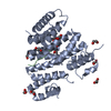
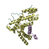
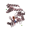
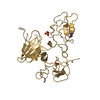
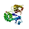
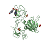
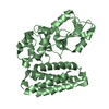
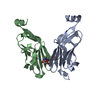
| Title | The crystal structure of plant specific calcium binding protein AtCBL2 in complex with the regulatory domain of AtCIPK14 | ||||||
 Components Components |
| ||||||
 Keywords Keywords |  SIGNALING PROTEIN/TRANSFERASE / calcium binding protein / protein-protein complex / ATP-binding / Kinase / Nucleotide-binding / Serine/threonine-protein kinase / Transferase / SIGNALING PROTEIN-TRANSFERASE COMPLEX SIGNALING PROTEIN/TRANSFERASE / calcium binding protein / protein-protein complex / ATP-binding / Kinase / Nucleotide-binding / Serine/threonine-protein kinase / Transferase / SIGNALING PROTEIN-TRANSFERASE COMPLEX | ||||||
| Function / homology |  Function and homology information Function and homology informationplant-type vacuole membrane / response to water deprivation / plasmodesma / response to abscisic acid / plant-type vacuole / potassium ion homeostasis / vacuole / plastid / response to glucose / cytosolic ribosome ...plant-type vacuole membrane / response to water deprivation / plasmodesma / response to abscisic acid / plant-type vacuole / potassium ion homeostasis / vacuole / plastid / response to glucose / cytosolic ribosome / response to salt stress / response to cold / calcium-mediated signaling / kinase binding / response to heat / response to oxidative stress / non-specific serine/threonine protein kinase / protein serine/threonine kinase activity / calcium ion binding / signal transduction / ATP binding / membrane / nucleus / cytoplasmSimilarity search - Function | ||||||
| Biological species |  Arabidopsis thaliana (thale cress) Arabidopsis thaliana (thale cress) | ||||||
| Method | X-RAY DIFFRACTION / SYNCHROTRON / MAD / Resolution: 1.2 Å | ||||||
 Authors Authors | Akaboshi, M. / Hashimoto, H. / Ishida, H. / Koizumi, N. / Sato, M. / Shimizu, T. | ||||||
 Citation Citation | Journal: J.Mol.Biol. / Year: 2008 Title: The crystal structure of plant-specific calcium-binding protein AtCBL2 in complex with the regulatory domain of AtCIPK14 Authors: Akaboshi, M. / Hashimoto, H. / Ishida, H. / Saijo, S. / Koizumi, N. / Sato, M. / Shimizu, T. | ||||||
| History |
|
- Structure visualization
| Structure viewer | Molecule: MolmilJmol/JSmol |
|---|
- Downloads & links
-Download
| PDBx/mmCIF format | 2zfd.cif.gz | 81 KB | Display | PDBx/mmCIF format |
|---|---|---|---|---|
| PDB format | pdb2zfd.ent.gz | 59 KB | Display | PDB format |
| PDBx/mmJSON format | 2zfd.json.gz | Tree view | PDBx/mmJSON format | |
| Others |  Other downloads Other downloads |
-Validation report
| Arichive directory | https://data.pdbj.org/pub/pdb/validation_reports/zf/2zfdftp://data.pdbj.org/pub/pdb/validation_reports/zf/2zfd | HTTPS FTP |
|---|
-Related structure data
| Related structure data | |
|---|---|
| Similar structure data |


-Links
 PDBj
PDBj


- Assembly
| Deposited unit | 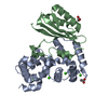
| ||||||||
|---|---|---|---|---|---|---|---|---|---|
| 1 |
| ||||||||
| Unit cell |
|
-Components
| #1: Protein | Mass: 25841.357 Da / Num. of mol.: 1 Source method: isolated from a genetically manipulated source Source: (gene. exp.) Arabidopsis thaliana (thale cress) / Gene: CBL2 / Plasmid: pETDuet-1 / Production host:  Escherichia coli (E. coli) / References: UniProt: Q8LAS7 Escherichia coli (E. coli) / References: UniProt: Q8LAS7 | ||||
|---|---|---|---|---|---|
| #2: Protein | Mass: 14055.048 Da / Num. of mol.: 1 / Fragment: UNP residues 305-427 Source method: isolated from a genetically manipulated source Source: (gene. exp.) Arabidopsis thaliana (thale cress) / Gene: CIPK14 / Plasmid: pETDuet-1 / Production host: Escherichia coli (E. coli) / References: UniProt: Q9LZW4 | ||||
| #3: Chemical | ChemComp-CA /   Mass: 40.078 Da / Num. of mol.: 6 / Source method: obtained synthetically / Formula: Ca Mass: 40.078 Da / Num. of mol.: 6 / Source method: obtained synthetically / Formula: Ca#4: Chemical | Acetic acid  Mass: 60.052 Da / Num. of mol.: 2 / Source method: obtained synthetically / Formula: C2H4O2 Mass: 60.052 Da / Num. of mol.: 2 / Source method: obtained synthetically / Formula: C2H4O2#5: Water | ChemComp-HOH / | Water Mass: 18.015 Da / Num. of mol.: 236 / Source method: isolated from a natural source / Formula: H2O Mass: 18.015 Da / Num. of mol.: 236 / Source method: isolated from a natural source / Formula: H2O |
-Experimental details
-Experiment
| Experiment | Method: X-RAY DIFFRACTION / Number of used crystals: 2 |
|---|
- Sample preparation
| Crystal | Density Matthews: 1.81 Å3/Da / Density % sol: 31.86 % |
|---|
-Data collection
| Diffraction source | Source: SYNCHROTRON / Site: Photon Factory  / Beamline: AR-NW12A / Wavelength: 0.9796, 0.9798, 0.9645 / Beamline: AR-NW12A / Wavelength: 0.9796, 0.9798, 0.9645 | ||||||||||||
|---|---|---|---|---|---|---|---|---|---|---|---|---|---|
| Detector | Type: ADSC QUANTUM 210 / Detector: CCD / Date: Feb 11, 2007 | ||||||||||||
| Radiation | Protocol: MAD / Monochromatic (M) / Laue (L): M / Scattering type: x-ray | ||||||||||||
| Radiation wavelength |
| ||||||||||||
| Reflection | Resolution: 1.2→50 Å / Num. obs: 85565 / % possible obs: 96.7 % / Observed criterion σ(F): 0 / Observed criterion σ(I): 0 / Redundancy: 3.8 % / Rmerge(I) obs: 0.058 | ||||||||||||
| Reflection shell | Resolution: 1.2→1.24 Å / Redundancy: 3.4 % / Rmerge(I) obs: 0.431 / % possible all: 91.3 |
- Processing
| Software |
| |||||||||||||||||||||||||||||||||||||||||||||||||||||||||||||||||||||||||||||||||||||||||||||||||||||||||||||||||||||||||||||||||||||||
|---|---|---|---|---|---|---|---|---|---|---|---|---|---|---|---|---|---|---|---|---|---|---|---|---|---|---|---|---|---|---|---|---|---|---|---|---|---|---|---|---|---|---|---|---|---|---|---|---|---|---|---|---|---|---|---|---|---|---|---|---|---|---|---|---|---|---|---|---|---|---|---|---|---|---|---|---|---|---|---|---|---|---|---|---|---|---|---|---|---|---|---|---|---|---|---|---|---|---|---|---|---|---|---|---|---|---|---|---|---|---|---|---|---|---|---|---|---|---|---|---|---|---|---|---|---|---|---|---|---|---|---|---|---|---|---|---|
| Refinement | Method to determine structure: MAD / Resolution: 1.2→20 Å / Cor.coef. Fo:Fc: 0.958 / Cor.coef. Fo:Fc free: 0.954 / SU B: 1.128 / SU ML: 0.024 / TLS residual ADP flag: LIKELY RESIDUAL / Cross valid method: THROUGHOUT / ESU R: 0.043 / ESU R Free: 0.043 / Stereochemistry target values: MAXIMUM LIKELIHOOD / Details: HYDROGENS HAVE BEEN ADDED IN THE RIDING POSITIONS
| |||||||||||||||||||||||||||||||||||||||||||||||||||||||||||||||||||||||||||||||||||||||||||||||||||||||||||||||||||||||||||||||||||||||
| Solvent computation | Ion probe radii: 0.8 Å / Shrinkage radii: 0.8 Å / VDW probe radii: 1.2 Å / Solvent model: BABINET MODEL WITH MASK | |||||||||||||||||||||||||||||||||||||||||||||||||||||||||||||||||||||||||||||||||||||||||||||||||||||||||||||||||||||||||||||||||||||||
| Displacement parameters | Biso mean: 7.746 Å2
| |||||||||||||||||||||||||||||||||||||||||||||||||||||||||||||||||||||||||||||||||||||||||||||||||||||||||||||||||||||||||||||||||||||||
| Refinement step | Cycle: LAST / Resolution: 1.2→20 Å
| |||||||||||||||||||||||||||||||||||||||||||||||||||||||||||||||||||||||||||||||||||||||||||||||||||||||||||||||||||||||||||||||||||||||
| Refine LS restraints |
| |||||||||||||||||||||||||||||||||||||||||||||||||||||||||||||||||||||||||||||||||||||||||||||||||||||||||||||||||||||||||||||||||||||||
| LS refinement shell | Resolution: 1.2→1.231 Å / Total num. of bins used: 20
| |||||||||||||||||||||||||||||||||||||||||||||||||||||||||||||||||||||||||||||||||||||||||||||||||||||||||||||||||||||||||||||||||||||||
| Refinement TLS params. | Method: refined / Origin x: 31.1778 Å / Origin y: 8.5398 Å / Origin z: 21.8775 Å
| |||||||||||||||||||||||||||||||||||||||||||||||||||||||||||||||||||||||||||||||||||||||||||||||||||||||||||||||||||||||||||||||||||||||
| Refinement TLS group |
|
