 Movie
Movie Controller

[English] 日本語
 Yorodumi
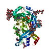
- PDB-2zal: Crystal structure of E. coli isoaspartyl aminopeptidase/L-asparag... -
+ Open data

- Basic information
| Entry | Database: PDB / ID: 2zal | |||||||||
|---|---|---|---|---|---|---|---|---|---|---|
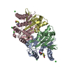
| Title | Crystal structure of E. coli isoaspartyl aminopeptidase/L-asparaginase in complex with L-aspartate | |||||||||
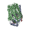
 Components Components | (L-asparaginase Asparaginase) x 2 Asparaginase) x 2 | |||||||||
 Keywords Keywords | HYDROLASE / isoaspartyl peptidase / asparaginase / Ntn-hydrolase / autoproteolysis / L-aspartate/calcium cluster | |||||||||
| Function / homology |  Function and homology informationbeta-aspartyl-peptidase / asparaginase activity / beta-aspartyl-peptidase activity / protein autoprocessing / hydrolase activity / cytoplasm Function and homology informationbeta-aspartyl-peptidase / asparaginase activity / beta-aspartyl-peptidase activity / protein autoprocessing / hydrolase activity / cytoplasmSimilarity search - Function | |||||||||
| Biological species |  Escherichia coli (E. coli) Escherichia coli (E. coli) | |||||||||
| Method | X-RAY DIFFRACTION / SYNCHROTRON / MOLECULAR REPLACEMENT / Resolution: 1.9 Å | |||||||||
 Authors Authors | Michalska, K. / Brzezinski, K. / Jaskolski, M. | |||||||||
 Citation Citation | Journal: J.Biol.Chem. / Year: 2005 Title: Crystal structure of isoaspartyl aminopeptidase in complex with L-aspartate Authors: Michalska, K. / Brzezinski, K. / Jaskolski, M. #1: Journal: Acta Crystallogr.,Sect.D / Year: 2000 Title: Crystallization and preliminary crystallographic studies of a new L-asparaginase encoded by the Escherichia coli genome Authors: Borek, D. / Jaskolski, M. #2: Journal: Nature / Year: 1995 Title: A protein catalytic framework with an N-terminal nucleophile is capable of self-activation Authors: Brannigan, J.A. / Dodson, G. / Duggleby, H.J. / Moody, P.C.E. / Smith, J.L. / Tomchick, D.R. / Murzin, A.G. #3: Journal: Protein Sci. / Year: 1998Title: Crystal structure of glycosylasparaginase from Flavobacterium meningosepticum Authors: Xuan, J. / Tarentino, A.L. / Grimwood, B.G. / Plummer Jr., T.H. / Cui, T. / Guan, C. / Van Roey, P. #4: Journal: Nat.Struct.Biol. / Year: 1995Title: Three-dimensional structure of human lysosomal aspartylglucosaminidase Authors: Oinonen, C. / Tikkanen, R. / Rouvinen, J. / Peltonen, L. #5: Journal: J.Biol.Chem. / Year: 1998Title: Crystal structures of Flavobacterium glycosylasparaginase. An N-terminal nucleophile hydrolase activated by intramolecular proteolysis Authors: Guo, H.-C. / Xu, Q. / Buckley, D. / Guan, C. #6: Journal: Cell(Cambridge,Mass.) / Year: 1999Title: Structural insights into the mechanism of intramolecular proteolysis Authors: Xu, Q. / Buckley, D. / Guan, C. / Guo, H.-C. #7: Journal: Biochem.J. / Year: 2004 Title: Autoproteolytic activation of human aspartylglucosaminidase Authors: Saarela, J. / Oinonen, C. / Jalanko, A. / Rouvinen, J. / Peltonen, L. #8: Journal: Acta Crystallogr.,Sect.D / Year: 2004Title: Structure of the isoaspartyl peptidase with L-asparaginase activity from Escherichia coli Authors: Prahl, A. / Pazgier, M. / Hejazi, M. / Lockau, W. / Lubkowski, J. | |||||||||
| History |
|
- Structure visualization
| Structure viewer | Molecule: MolmilJmol/JSmol |
|---|
- Downloads & links
-Download
| PDBx/mmCIF format | 2zal.cif.gz | 128.5 KB | Display | PDBx/mmCIF format |
|---|---|---|---|---|
| PDB format | pdb2zal.ent.gz | 99.7 KB | Display | PDB format |
| PDBx/mmJSON format | 2zal.json.gz | Tree view | PDBx/mmJSON format | |
| Others |  Other downloads Other downloads |
-Validation report
| Arichive directory | https://data.pdbj.org/pub/pdb/validation_reports/za/2zalftp://data.pdbj.org/pub/pdb/validation_reports/za/2zal | HTTPS FTP |
|---|
-Related structure data
| Related structure data |  1k2xS S: Starting model for refinement |
|---|---|
| Similar structure data |


-Links
 PDBj
PDBj


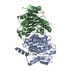
- Assembly
| Deposited unit | 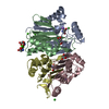
| ||||||||
|---|---|---|---|---|---|---|---|---|---|
| 1 |
| ||||||||
| Unit cell |
|
-Components
-Protein , 2 types, 4 molecules ACBD
| #1: Protein | Asparaginase / L-ASPARAGINASE SUBUNIT ALPHA / L-asparagine amidohydrolase Mass: 17071.527 Da / Num. of mol.: 2 / Fragment: N-terminal subunit (alpha), UNP residues 2-161 Source method: isolated from a genetically manipulated source Source: (gene. exp.) Escherichia coli (E. coli) / Strain: K12Description: The N-terminal methionine has been removed by an intracellular aminopeptidase (E. coli). During expression and purification, the protein undergoes autoproteolytic cleavage into alpha and ...Description: The N-terminal methionine has been removed by an intracellular aminopeptidase (E. coli). During expression and purification, the protein undergoes autoproteolytic cleavage into alpha and beta subunits. The residues 162-178 were not present in the crystallized material due to partial, non-specific degradation at the c-terminus. The number of missing residues was determined by mass spectrometry Gene: ybiK (iaaA) / Plasmid: pET11d / Production host: Escherichia coli (E. coli) / Strain (production host): BL21(DE3)pLysSReferences: UniProt: P37595, beta-aspartyl-peptidase, asparaginase#2: Protein | Asparaginase / L-ASPARAGINASE SUBUNIT BETA / L-asparagine amidohydrolaseMass: 13812.512 Da / Num. of mol.: 2 / Fragment: C-terminal subunit (beta), UNP residues 179-315 Source method: isolated from a genetically manipulated source Source: (gene. exp.) Escherichia coli (E. coli) / Strain: K12Description: During expression and purification, the protein undergoes autoproteolytic cleavage into alpha and beta subunits. The residues 316-321 were not present in the crystallized material due to ...Description: During expression and purification, the protein undergoes autoproteolytic cleavage into alpha and beta subunits. The residues 316-321 were not present in the crystallized material due to partial, non-specific degradation at the C-terminus. The number of missing residues was determined by mass spectrometry Gene: ybiK (iaaA) / Plasmid: pET11d / Production host: Escherichia coli (E. coli) / Strain (production host): BL21(DE3)pLysSReferences: UniProt: P37595, beta-aspartyl-peptidase, asparaginase |
|---|
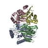
-Non-polymers , 6 types, 263 molecules 


| #3: Chemical |  Mass: 22.990 Da / Num. of mol.: 2 / Source method: obtained synthetically / Formula: Na Mass: 22.990 Da / Num. of mol.: 2 / Source method: obtained synthetically / Formula: Na#4: Chemical | ChemComp-CA /  Mass: 40.078 Da / Num. of mol.: 5 / Source method: obtained synthetically / Formula: Ca Mass: 40.078 Da / Num. of mol.: 5 / Source method: obtained synthetically / Formula: Ca#5: Chemical | ChemComp-ASP / Aspartic acid Type: L-peptide linking / Mass: 133.103 Da / Num. of mol.: 5 / Source method: obtained synthetically / Formula: C4H7NO4 Type: L-peptide linking / Mass: 133.103 Da / Num. of mol.: 5 / Source method: obtained synthetically / Formula: C4H7NO4#6: Chemical | ChemComp-CL / | Chloride Mass: 35.453 Da / Num. of mol.: 1 / Source method: obtained synthetically / Formula: Cl Mass: 35.453 Da / Num. of mol.: 1 / Source method: obtained synthetically / Formula: Cl#7: Chemical | Tris Mass: 122.143 Da / Num. of mol.: 2 / Source method: obtained synthetically / Formula: C4H12NO3 / Comment: pH buffer*YM Mass: 122.143 Da / Num. of mol.: 2 / Source method: obtained synthetically / Formula: C4H12NO3 / Comment: pH buffer*YM#8: Water | ChemComp-HOH / | WaterMass: 18.015 Da / Num. of mol.: 248 / Source method: isolated from a natural source / Formula: H2O |
|---|
-Experimental details
-Experiment
| Experiment | Method: X-RAY DIFFRACTION / Number of used crystals: 1 |
|---|
- Sample preparation
| Crystal | Density Matthews: 2.3 Å3/Da / Density % sol: 46.41 % |
|---|---|
| Crystal grow | Temperature: 292 K / Method: vapor diffusion, hanging drop / pH: 8.5 Details: 100mM Tris/HCl, 80mM calcium chloride, 100mM sodium aspartate, 17% PEG 4000, 13% PEG 400, pH 8.5, VAPOR DIFFUSION, HANGING DROP, temperature 292K |
-Data collection
| Diffraction | Mean temperature: 100 K |
|---|---|
| Diffraction source | Source: SYNCHROTRON / Site: MAX II  / Beamline: I711 / Wavelength: 1.095 Å / Beamline: I711 / Wavelength: 1.095 Å |
| Detector | Type: MARRESEARCH / Detector: CCD / Date: Apr 28, 2002 |
| Radiation | Monochromator: Si single crystal / Protocol: SINGLE WAVELENGTH / Monochromatic (M) / Laue (L): M / Scattering type: x-ray |
| Radiation wavelength | Wavelength: 1.095 Å / Relative weight: 1 |
| Reflection | Resolution: 1.9→20 Å / Num. all: 43572 / Num. obs: 43572 / % possible obs: 95.8 % / Observed criterion σ(F): 0 / Observed criterion σ(I): -3 / Redundancy: 3.5 % / Biso Wilson estimate: 18.67 Å2 / Rmerge(I) obs: 0.064 / Net I/σ(I): 15 |
| Reflection shell | Resolution: 1.9→1.97 Å / Redundancy: 3 % / Rmerge(I) obs: 0.256 / Mean I/σ(I) obs: 2.2 / Num. unique all: 4085 / % possible all: 91.3 |
- Processing
| Software |
| |||||||||||||||||||||||||||||||||||||||||||||||||||||||||||||||||||||||||||||||||||||||||||||||||||||||||||||||||||||||||||||
|---|---|---|---|---|---|---|---|---|---|---|---|---|---|---|---|---|---|---|---|---|---|---|---|---|---|---|---|---|---|---|---|---|---|---|---|---|---|---|---|---|---|---|---|---|---|---|---|---|---|---|---|---|---|---|---|---|---|---|---|---|---|---|---|---|---|---|---|---|---|---|---|---|---|---|---|---|---|---|---|---|---|---|---|---|---|---|---|---|---|---|---|---|---|---|---|---|---|---|---|---|---|---|---|---|---|---|---|---|---|---|---|---|---|---|---|---|---|---|---|---|---|---|---|---|---|---|
| Refinement | Method to determine structure: MOLECULAR REPLACEMENT Starting model: PDB entry 1K2X Resolution: 1.9→20 Å / Cor.coef. Fo:Fc: 0.965 / Cor.coef. Fo:Fc free: 0.955 / SU B: 2.746 / SU ML: 0.08 / TLS residual ADP flag: LIKELY RESIDUAL / Isotropic thermal model: Isotropic / Cross valid method: R-free / σ(F): 0 / σ(I): 0 / ESU R: 0.135 / ESU R Free: 0.121 / Stereochemistry target values: Engh & Huber Details: MAXIMUM LIKELIHOOD TARGET. THE REFINEMENT INCLUDED TLS PARAMETERS. THE RESIDUES 158-161 FROM CHAIN C (SUBUNIT ALPHA) AS WELL AS THE RESIDUES 314-315 FROM THE CHAINS B AND D (SUBUNITS BETA) ...Details: MAXIMUM LIKELIHOOD TARGET. THE REFINEMENT INCLUDED TLS PARAMETERS. THE RESIDUES 158-161 FROM CHAIN C (SUBUNIT ALPHA) AS WELL AS THE RESIDUES 314-315 FROM THE CHAINS B AND D (SUBUNITS BETA) WERE NOT MODELED DUE TO POOR ELECTRON DENSITY. CHAIN A IS COMPLETE. IN EACH OF THE TWO ACTIVE SITES, A CLEARLY VISIBLE PRODUCT OF THE ENZYMATIC REACTION, L-ASPARTATE, IS MODELED. IN INTERSTITIAL POSITIONS, THE STRUCTURE CONTAINS A BIG 3L-ASP/5CA COORDINATION COMPLEX.
| |||||||||||||||||||||||||||||||||||||||||||||||||||||||||||||||||||||||||||||||||||||||||||||||||||||||||||||||||||||||||||||
| Solvent computation | Ion probe radii: 0.8 Å / Shrinkage radii: 0.8 Å / VDW probe radii: 1.4 Å / Solvent model: BABINET MODEL WITH MASK | |||||||||||||||||||||||||||||||||||||||||||||||||||||||||||||||||||||||||||||||||||||||||||||||||||||||||||||||||||||||||||||
| Displacement parameters | Biso mean: 13.27 Å2
| |||||||||||||||||||||||||||||||||||||||||||||||||||||||||||||||||||||||||||||||||||||||||||||||||||||||||||||||||||||||||||||
| Refinement step | Cycle: LAST / Resolution: 1.9→20 Å
| |||||||||||||||||||||||||||||||||||||||||||||||||||||||||||||||||||||||||||||||||||||||||||||||||||||||||||||||||||||||||||||
| Refine LS restraints |
| |||||||||||||||||||||||||||||||||||||||||||||||||||||||||||||||||||||||||||||||||||||||||||||||||||||||||||||||||||||||||||||
| LS refinement shell | Resolution: 1.904→2.006 Å / Total num. of bins used: 10 /
| |||||||||||||||||||||||||||||||||||||||||||||||||||||||||||||||||||||||||||||||||||||||||||||||||||||||||||||||||||||||||||||
| Refinement TLS params. | Method: refined / Refine-ID: X-RAY DIFFRACTION
| |||||||||||||||||||||||||||||||||||||||||||||||||||||||||||||||||||||||||||||||||||||||||||||||||||||||||||||||||||||||||||||
| Refinement TLS group |
|
