 ムービー
ムービー コントローラー


+ データを開く

- 基本情報
基本情報
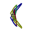
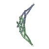
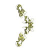
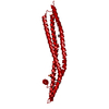
| 登録情報 | データベース: PDB / ID: 2z0n | ||||||
|---|---|---|---|---|---|---|---|
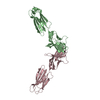
| タイトル | Crystal structure of APPL1-BAR domain | ||||||
 要素 要素 | DCC-interacting protein 13-alpha | ||||||
 キーワード キーワード |  SIGNALING PROTEIN / helix bundle (ヘリックスバンドル) / Cell cycle (細胞周期) / Coiled coil (コイルドコイル) / Endosome (エンドソーム) / Membrane (生体膜) / Nucleus (Nucleus) / Phosphorylation (リン酸化) / Structural Genomics (構造ゲノミクス) / NPPSFA / National Project on Protein Structural and Functional Analyses / RIKEN Structural Genomics/Proteomics Initiative / RSGI SIGNALING PROTEIN / helix bundle (ヘリックスバンドル) / Cell cycle (細胞周期) / Coiled coil (コイルドコイル) / Endosome (エンドソーム) / Membrane (生体膜) / Nucleus (Nucleus) / Phosphorylation (リン酸化) / Structural Genomics (構造ゲノミクス) / NPPSFA / National Project on Protein Structural and Functional Analyses / RIKEN Structural Genomics/Proteomics Initiative / RSGI | ||||||
| 機能・相同性 |  機能・相同性情報 機能・相同性情報negative regulation of Fc-gamma receptor signaling pathway involved in phagocytosis / positive regulation of macropinocytosis / adiponectin-activated signaling pathway / マクロピノソーム / regulation of fibroblast migration / regulation of glucose import / maintenance of synapse structure / protein kinase B binding / signaling / positive regulation of melanin biosynthetic process ...negative regulation of Fc-gamma receptor signaling pathway involved in phagocytosis / positive regulation of macropinocytosis / adiponectin-activated signaling pathway / マクロピノソーム / regulation of fibroblast migration / regulation of glucose import / maintenance of synapse structure / protein kinase B binding / signaling / positive regulation of melanin biosynthetic process / regulation of toll-like receptor 4 signaling pathway / vesicle membrane / early phagosome / Caspase activation via Dependence Receptors in the absence of ligand / positive regulation of cytokine production involved in inflammatory response / cellular response to hepatocyte growth factor stimulus / intracellular vesicle / regulation of innate immune response / beta-tubulin binding / phosphatidylserine binding / regulation of G1/S transition of mitotic cell cycle / regulation of protein localization to plasma membrane / ruffle / phosphatidylinositol binding / transforming growth factor beta receptor signaling pathway / positive regulation of glucose import / protein import into nucleus / presynapse / insulin receptor signaling pathway / early endosome membrane / cytoplasmic vesicle / postsynapse / エンドソーム / endosome membrane / エンドソーム / 細胞周期 / glutamatergic synapse / protein-containing complex binding / シグナル伝達 / protein homodimerization activity / extracellular exosome / 生体膜 / identical protein binding / 細胞核 / 細胞膜 / 細胞質基質 / 細胞質類似検索 - 分子機能 | ||||||
| 生物種 |  Homo sapiens (ヒト) Homo sapiens (ヒト) | ||||||
| 手法 | X線回折 / シンクロトロン / 多波長異常分散 / 解像度: 1.95 Å | ||||||
 データ登録者 データ登録者 | Murayama, K. / Kato-Murayama, M. / Sakamoto, A. / Shirouzu, M. / Yokoyama, S. / RIKEN Structural Genomics/Proteomics Initiative (RSGI) | ||||||
 引用 引用 | ジャーナル: To be Published タイトル: Crystal structure of APPL1-BAR domain 著者: Murayama, K. / Kato-Murayama, M. / Sakamoto, A. / Shirouzu, M. / Yokoyama, S. | ||||||
| 履歴 |
|
- 構造の表示
構造の表示
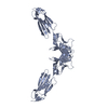
| 構造ビューア | 分子: MolmilJmol/JSmol |
|---|
- ダウンロードとリンク
-ダウンロード
| PDBx/mmCIF形式 | 2z0n.cif.gz | 59.1 KB | 表示 | PDBx/mmCIF形式 |
|---|---|---|---|---|
| PDB形式 | pdb2z0n.ent.gz | 46.7 KB | 表示 | PDB形式 |
| PDBx/mmJSON形式 | 2z0n.json.gz | ツリー表示 | PDBx/mmJSON形式 | |
| その他 |  その他のダウンロード その他のダウンロード |
-検証レポート
| アーカイブディレクトリ | https://data.pdbj.org/pub/pdb/validation_reports/z0/2z0nftp://data.pdbj.org/pub/pdb/validation_reports/z0/2z0n | HTTPS FTP |
|---|
-関連構造データ
| 類似構造データ | |
|---|---|
| その他のデータベース |

-リンク
 PDBj
PDBj


- 集合体
集合体
| 登録構造単位 | 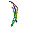
| ||||||||
|---|---|---|---|---|---|---|---|---|---|
| 1 | 
| ||||||||
| 単位格子 |
|
-要素
| #1: タンパク質 | 分子量: 32708.053 Da / 分子数: 1 / 断片: reisdues 1-275 / 由来タイプ: 組換発現 / 由来: (組換発現) Homo sapiens (ヒト) / プラスミド: pGEX / 発現宿主:  Escherichia coli (大腸菌) / 参照: UniProt: Q9UKG1 Escherichia coli (大腸菌) / 参照: UniProt: Q9UKG1 |
|---|---|
| #2: 水 | ChemComp-HOH / 水 分子量: 18.015 Da / 分子数: 96 / 由来タイプ: 天然 / 式: H2O 分子量: 18.015 Da / 分子数: 96 / 由来タイプ: 天然 / 式: H2O |
-実験情報
-実験
| 実験 | 手法: X線回折 / 使用した結晶の数: 1 |
|---|
- 試料調製
試料調製
| 結晶 | マシュー密度: 1.96 Å3/Da / 溶媒含有率: 37.32 % |
|---|---|
| 結晶化 | 温度: 293 K / 手法: 蒸気拡散法, ハンギングドロップ法 / pH: 7.5 詳細: 17.5% PEG3350, 0.2M Ammonium sulfate, 0.1M Hepes, VAPOR DIFFUSION, HANGING DROP, temperature 293K |
-データ収集
| 回折 | 平均測定温度: 100 K | ||||||||||||
|---|---|---|---|---|---|---|---|---|---|---|---|---|---|
| 放射光源 | 由来: シンクロトロン / サイト: SPring-8  / ビームライン: BL44B2 / 波長: 0.979089, 0.979462, 0.964 / ビームライン: BL44B2 / 波長: 0.979089, 0.979462, 0.964 | ||||||||||||
| 検出器 | タイプ: ADSC QUANTUM 210 / 検出器: CCD / 日付: 2006年9月26日 | ||||||||||||
| 放射 | プロトコル: MAD / 単色(M)・ラウエ(L): M / 散乱光タイプ: x-ray | ||||||||||||
| 放射波長 |
| ||||||||||||
| 反射 | 解像度: 1.95→50 Å / Num. obs: 19533 / % possible obs: 99.9 % / Observed criterion σ(F): -3 / 冗長度: 7.1 % / Biso Wilson estimate: 19.1 Å2 / Rsym value: 0.08 / Net I/σ(I): 16.8 | ||||||||||||
| 反射 シェル | 解像度: 1.95→2.02 Å / Rsym value: 0.341 / % possible all: 100 |
- 解析
解析
| ソフトウェア |
| ||||||||||||||||||||||||||||||||||||||||||||||||||||||||||||||||||||||||||||||||
|---|---|---|---|---|---|---|---|---|---|---|---|---|---|---|---|---|---|---|---|---|---|---|---|---|---|---|---|---|---|---|---|---|---|---|---|---|---|---|---|---|---|---|---|---|---|---|---|---|---|---|---|---|---|---|---|---|---|---|---|---|---|---|---|---|---|---|---|---|---|---|---|---|---|---|---|---|---|---|---|---|---|
| 精密化 | 構造決定の手法: 多波長異常分散 / 解像度: 1.95→35.52 Å / Rfactor Rfree error: 0.006 / Data cutoff high absF: 1385109.6 / Data cutoff low absF: 0 / Isotropic thermal model: RESTRAINED / 交差検証法: THROUGHOUT / σ(F): 0
| ||||||||||||||||||||||||||||||||||||||||||||||||||||||||||||||||||||||||||||||||
| 溶媒の処理 | 溶媒モデル: FLAT MODEL / Bsol: 43.905 Å2 / ksol: 0.375848 e/Å3 | ||||||||||||||||||||||||||||||||||||||||||||||||||||||||||||||||||||||||||||||||
| 原子変位パラメータ | Biso mean: 31.2 Å2
| ||||||||||||||||||||||||||||||||||||||||||||||||||||||||||||||||||||||||||||||||
| Refine analyze |
| ||||||||||||||||||||||||||||||||||||||||||||||||||||||||||||||||||||||||||||||||
| 精密化ステップ | サイクル: LAST / 解像度: 1.95→35.52 Å
| ||||||||||||||||||||||||||||||||||||||||||||||||||||||||||||||||||||||||||||||||
| 拘束条件 |
| ||||||||||||||||||||||||||||||||||||||||||||||||||||||||||||||||||||||||||||||||
| LS精密化 シェル | 解像度: 1.95→2.07 Å / Rfactor Rfree error: 0.016 / Total num. of bins used: 6
| ||||||||||||||||||||||||||||||||||||||||||||||||||||||||||||||||||||||||||||||||
| Xplor file |
|
