 Movie
Movie Controller


+ Open data

- Basic information
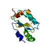
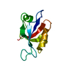
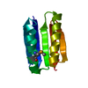
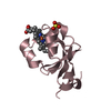
| Entry | Database: PDB / ID: 2yvi | ||||||
|---|---|---|---|---|---|---|---|
| Title | Crystal structure of a death domain of human ankryn protein | ||||||
 Components Components | Ankyrin-1 | ||||||
 Keywords Keywords | CELL ADHESION / homo sapiens / death domain / monomer / Structural Genomics / NPPSFA / National Project on Protein Structural and Functional Analyses / RIKEN Structural Genomics/Proteomics Initiative / RSGI | ||||||
| Function / homology |  Function and homology information Function and homology informationspectrin-associated cytoskeleton / positive regulation of organelle organization / maintenance of epithelial cell apical/basal polarity / NrCAM interactions / Neurofascin interactions / ankyrin-1 complex / CHL1 interactions / cytoskeletal anchor activity / M band / Interaction between L1 and Ankyrins ...spectrin-associated cytoskeleton / positive regulation of organelle organization / maintenance of epithelial cell apical/basal polarity / NrCAM interactions / Neurofascin interactions / ankyrin-1 complex / CHL1 interactions / cytoskeletal anchor activity / M band / Interaction between L1 and Ankyrins / spectrin binding / exocytosis / axolemma / endoplasmic reticulum to Golgi vesicle-mediated transport / COPI-mediated anterograde transport / cytoskeleton organization / sarcoplasmic reticulum / protein localization to plasma membrane / sarcolemma / cytoplasmic side of plasma membrane / structural constituent of cytoskeleton / Z disc / ATPase binding / postsynaptic membrane / basolateral plasma membrane / protein phosphatase binding / transmembrane transporter binding / cytoskeleton / neuron projection / structural molecule activity / enzyme binding / signal transduction / plasma membrane / cytosolSimilarity search - Function | ||||||
| Biological species |  Homo sapiens (human) Homo sapiens (human) | ||||||
| Method | X-RAY DIFFRACTION / SYNCHROTRON / SAD / Resolution: 1.92 Å | ||||||
 Authors Authors | Ihsanawati / Bessho, Y. / Chen, L. / Liu, Z.J. / Wang, B.C. / Shirouzu, M. / Yokoyama, S. / RIKEN Structural Genomics/Proteomics Initiative (RSGI) | ||||||
 Citation Citation | Journal: To be Published Title: Crystal structure of a death domain of human ankryn protein Authors: Ihsanawati / Bessho, Y. / Chen, L. / Liu, Z.J. / Wang, B.C. / Shirouzu, M. / Yokoyama, S. | ||||||
| History |
|
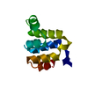
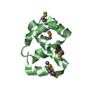
- Structure visualization
| Structure viewer | Molecule: MolmilJmol/JSmol |
|---|
- Downloads & links
-Download
| PDBx/mmCIF format | 2yvi.cif.gz | 32.5 KB | Display | PDBx/mmCIF format |
|---|---|---|---|---|
| PDB format | pdb2yvi.ent.gz | 21.4 KB | Display | PDB format |
| PDBx/mmJSON format | 2yvi.json.gz | Tree view | PDBx/mmJSON format | |
| Others |  Other downloads Other downloads |
-Validation report
| Arichive directory | https://data.pdbj.org/pub/pdb/validation_reports/yv/2yviftp://data.pdbj.org/pub/pdb/validation_reports/yv/2yvi | HTTPS FTP |
|---|
-Related structure data
| Similar structure data | |
|---|---|
| Other databases |


-Links
 PDBj
PDBj


- Assembly
| Deposited unit | 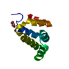
| ||||||||
|---|---|---|---|---|---|---|---|---|---|
| 1 |
| ||||||||
| 2 | 
| ||||||||
| Unit cell |
|
-Components
| #1: Protein | / Erythrocyte ankyrin / Ankyrin-R / death domain Mass: 12208.591 Da / Num. of mol.: 1 / Fragment: UNP residues 1394-1497, death domain Source method: isolated from a genetically manipulated source Source: (gene. exp.) Homo sapiens (human) / Description: Cell Free system / Gene: ANK1, ANK / Plasmid: PK060123-20 / References: UniProt: P16157 |
|---|---|
| #2: Chemical | ChemComp-GOL / Glycerol  Mass: 92.094 Da / Num. of mol.: 1 / Source method: obtained synthetically / Formula: C3H8O3 Mass: 92.094 Da / Num. of mol.: 1 / Source method: obtained synthetically / Formula: C3H8O3 |
| #3: Water | ChemComp-HOH / Water Mass: 18.015 Da / Num. of mol.: 94 / Source method: isolated from a natural source / Formula: H2O Mass: 18.015 Da / Num. of mol.: 94 / Source method: isolated from a natural source / Formula: H2O |
-Experimental details
-Experiment
| Experiment | Method: X-RAY DIFFRACTION / Number of used crystals: 1 |
|---|
- Sample preparation
| Crystal | Density Matthews: 1.87 Å3/Da / Density % sol: 34.05 % |
|---|---|
| Crystal grow | Temperature: 298 K / Method: vapor diffusion, sitting drop / pH: 8.5 Details: 0.2M NaCl, 0.1M TrisCl pH8.5, 25% PEG3350, VAPOR DIFFUSION, SITTING DROP, temperature 298K |
-Data collection
| Diffraction | Mean temperature: 100 K |
|---|---|
| Diffraction source | Source: SYNCHROTRON / Site: APS  / Beamline: 22-BM / Wavelength: 0.9782 Å / Beamline: 22-BM / Wavelength: 0.9782 Å |
| Detector | Type: MARMOSAIC 225 mm CCD / Detector: CCD / Date: Dec 18, 2006 |
| Radiation | Monochromator: Si / Protocol: MAD / Monochromatic (M) / Laue (L): M / Scattering type: x-ray |
| Radiation wavelength | Wavelength: 0.9782 Å / Relative weight: 1 |
| Reflection | Resolution: 1.92→30 Å / Num. obs: 7800 / % possible obs: 99.8 % / Observed criterion σ(I): -3 / Redundancy: 30.4 % / Biso Wilson estimate: 16.5 Å2 / Rsym value: 0.087 |
| Reflection shell | Resolution: 1.92→1.99 Å / Rmerge(I) obs: 0.187 / % possible all: 99.9 |
- Processing
| Software |
| ||||||||||||||||||||
|---|---|---|---|---|---|---|---|---|---|---|---|---|---|---|---|---|---|---|---|---|---|
| Refinement | Method to determine structure: SAD / Resolution: 1.92→26.99 Å / Rfactor Rfree error: 0.011 / Data cutoff high absF: 4857158.22 / Data cutoff low absF: 0 / Isotropic thermal model: RESTRAINED / Cross valid method: THROUGHOUT / σ(F): 0 / Stereochemistry target values: Engh & Huber
| ||||||||||||||||||||
| Solvent computation | Solvent model: FLAT MODEL / Bsol: 42.9707 Å2 / ksol: 0.383766 e/Å3 | ||||||||||||||||||||
| Displacement parameters | Biso mean: 22.1 Å2
| ||||||||||||||||||||
| Refine analyze |
| ||||||||||||||||||||
| Refinement step | Cycle: LAST / Resolution: 1.92→26.99 Å
| ||||||||||||||||||||
| Refine LS restraints |
| ||||||||||||||||||||
| LS refinement shell | Resolution: 1.9→2.02 Å / Rfactor Rfree error: 0.04 / Total num. of bins used: 6
| ||||||||||||||||||||
| Xplor file |
|
