 Movie
Movie Controller

[English] 日本語
 Yorodumi
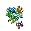
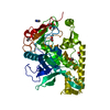
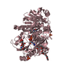
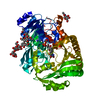
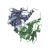
- PDB-2ys6: Crystal structure of GAR synthetase from Geobacillus kaustophilus -
+ Open data

- Basic information
| Entry | Database: PDB / ID: 2ys6 | ||||||
|---|---|---|---|---|---|---|---|
| Title | Crystal structure of GAR synthetase from Geobacillus kaustophilus | ||||||
 Components Components | Phosphoribosylglycinamide synthetase | ||||||
 Keywords Keywords |  LIGASE / Glycinamide ribonucleotide synthetase / GAR synthetase / ATP binding / Purine nucleotide biosynthetic pathway / Structural Genomics / NPPSFA / National Project on Protein Structural and Functional Analyses / RIKEN Structural Genomics/Proteomics Initiative / RSGI LIGASE / Glycinamide ribonucleotide synthetase / GAR synthetase / ATP binding / Purine nucleotide biosynthetic pathway / Structural Genomics / NPPSFA / National Project on Protein Structural and Functional Analyses / RIKEN Structural Genomics/Proteomics Initiative / RSGI | ||||||
| Function / homology |  Function and homology informationphosphoribosylamine-glycine ligase / phosphoribosylamine-glycine ligase activity / purine nucleobase biosynthetic process / 'de novo' IMP biosynthetic process / ATP binding / metal ion binding Function and homology informationphosphoribosylamine-glycine ligase / phosphoribosylamine-glycine ligase activity / purine nucleobase biosynthetic process / 'de novo' IMP biosynthetic process / ATP binding / metal ion bindingSimilarity search - Function | ||||||
| Biological species |  Geobacillus kaustophilus (bacteria) Geobacillus kaustophilus (bacteria) | ||||||
| Method | X-RAY DIFFRACTION / SYNCHROTRON / MOLECULAR REPLACEMENT / Resolution: 2.21 Å | ||||||
 Authors Authors | Baba, S. / Kanagawa, M. / Kuramitsu, S. / Yokoyama, S. / Kawai, G. / Sampei, G. / RIKEN Structural Genomics/Proteomics Initiative (RSGI) | ||||||
 Citation Citation | Journal: J.Biochem. / Year: 2010 Title: Crystal structures of glycinamide ribonucleotide synthetase, PurD, from thermophilic eubacteria Authors: Sampei, G. / Baba, S. / Kanagawa, M. / Yanai, H. / Ishii, T. / Kawai, H. / Fukai, Y. / Ebihara, A. / Nakagawa, N. / Kawai, G. | ||||||
| History |
|
- Structure visualization
| Structure viewer | Molecule: MolmilJmol/JSmol |
|---|
- Downloads & links
-Download
| PDBx/mmCIF format | 2ys6.cif.gz | 96.6 KB | Display | PDBx/mmCIF format |
|---|---|---|---|---|
| PDB format | pdb2ys6.ent.gz | 71.2 KB | Display | PDB format |
| PDBx/mmJSON format | 2ys6.json.gz | Tree view | PDBx/mmJSON format | |
| Others |  Other downloads Other downloads |
-Validation report
| Arichive directory | https://data.pdbj.org/pub/pdb/validation_reports/ys/2ys6ftp://data.pdbj.org/pub/pdb/validation_reports/ys/2ys6 | HTTPS FTP |
|---|
-Related structure data
| Related structure data | 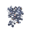 2ip4C  2yrwSC 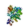 2yrxC  2ys7C 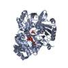 2yw2C 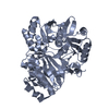 2yyaC S: Starting model for refinement C: citing same article ( |
|---|---|
| Similar structure data | |
| Other databases |


-Links
 PDBj
PDBj


- Assembly
| Deposited unit | 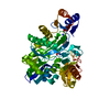
| ||||||||
|---|---|---|---|---|---|---|---|---|---|
| 1 |
| ||||||||
| Unit cell |
|
-Components
| #1: Protein | Mass: 48228.781 Da / Num. of mol.: 1 Source method: isolated from a genetically manipulated source Source: (gene. exp.) Geobacillus kaustophilus (bacteria) / Plasmid: pET-HisTEV / Production host: Escherichia coli (E. coli)References: UniProt: Q5L3C7, phosphoribosylamine-glycine ligase |
|---|---|
| #2: Chemical | ChemComp-GLY / Glycine  Type: peptide linking / Mass: 75.067 Da / Num. of mol.: 1 / Source method: obtained synthetically / Formula: C2H5NO2 Type: peptide linking / Mass: 75.067 Da / Num. of mol.: 1 / Source method: obtained synthetically / Formula: C2H5NO2 |
| #3: Chemical | ChemComp-AMP / Adenosine monophosphate  Mass: 347.221 Da / Num. of mol.: 1 / Source method: obtained synthetically / Formula: C10H14N5O7P / Comment: AMP*YM Mass: 347.221 Da / Num. of mol.: 1 / Source method: obtained synthetically / Formula: C10H14N5O7P / Comment: AMP*YM |
| #4: Water | ChemComp-HOH / Water Mass: 18.015 Da / Num. of mol.: 82 / Source method: isolated from a natural source / Formula: H2O Mass: 18.015 Da / Num. of mol.: 82 / Source method: isolated from a natural source / Formula: H2O |
-Experimental details
-Experiment
| Experiment | Method: X-RAY DIFFRACTION / Number of used crystals: 1 |
|---|
- Sample preparation
| Crystal | Density Matthews: 2.12 Å3/Da / Density % sol: 42 % |
|---|---|
| Crystal grow | Temperature: 293 K / Method: vapor diffusion, sitting drop / pH: 4.5 Details: 0.2M sodium dihydrogen phosphate monohydrate, 20%w/v polyethylene glycol 3350, 10mM ATP, 10mM Gly, pH 4.5, VAPOR DIFFUSION, SITTING DROP, temperature 293K |
-Data collection
| Diffraction | Mean temperature: 100 K |
|---|---|
| Diffraction source | Source: SYNCHROTRON / Site: SPring-8  / Beamline: BL26B2 / Wavelength: 1 Å / Beamline: BL26B2 / Wavelength: 1 Å |
| Detector | Type: RIGAKU JUPITER 210 / Detector: CCD / Date: Dec 15, 2006 |
| Radiation | Monochromator: Fixed exit Si 111 double crystal monochromator Protocol: SINGLE WAVELENGTH / Monochromatic (M) / Laue (L): M / Scattering type: x-ray |
| Radiation wavelength | Wavelength: 1 Å / Relative weight: 1 |
| Reflection | Resolution: 2.2→50 Å / Num. obs: 21104 / % possible obs: 99.7 % / Redundancy: 6.8 % / Biso Wilson estimate: 15 Å2 / Rmerge(I) obs: 0.089 |
| Reflection shell | Resolution: 2.2→2.28 Å / Redundancy: 6.3 % / Rmerge(I) obs: 0.424 / Num. unique all: 2041 / % possible all: 98.6 |
- Processing
| Software |
| ||||||||||||||||||||||||||||||||||||||||||||||||||||||||||||
|---|---|---|---|---|---|---|---|---|---|---|---|---|---|---|---|---|---|---|---|---|---|---|---|---|---|---|---|---|---|---|---|---|---|---|---|---|---|---|---|---|---|---|---|---|---|---|---|---|---|---|---|---|---|---|---|---|---|---|---|---|---|
| Refinement | Method to determine structure: MOLECULAR REPLACEMENT Starting model: PDB ENTRY 2YRW Resolution: 2.21→38.29 Å / Rfactor Rfree error: 0.005 / Data cutoff high absF: 72267.98 / Data cutoff low absF: 0 / Isotropic thermal model: RESTRAINED / Cross valid method: THROUGHOUT / σ(F): 0 / Stereochemistry target values: MAXIMUM LIKELIHOOD
| ||||||||||||||||||||||||||||||||||||||||||||||||||||||||||||
| Solvent computation | Solvent model: FLAT MODEL / Bsol: 38.5589 Å2 / ksol: 0.37007 e/Å3 | ||||||||||||||||||||||||||||||||||||||||||||||||||||||||||||
| Displacement parameters | Biso mean: 27 Å2
| ||||||||||||||||||||||||||||||||||||||||||||||||||||||||||||
| Refine analyze |
| ||||||||||||||||||||||||||||||||||||||||||||||||||||||||||||
| Refinement step | Cycle: LAST / Resolution: 2.21→38.29 Å
| ||||||||||||||||||||||||||||||||||||||||||||||||||||||||||||
| Refine LS restraints |
| ||||||||||||||||||||||||||||||||||||||||||||||||||||||||||||
| LS refinement shell | Resolution: 2.2→2.34 Å / Rfactor Rfree error: 0.016 / Total num. of bins used: 6
| ||||||||||||||||||||||||||||||||||||||||||||||||||||||||||||
| Xplor file |
|
