 Movie
Movie Controller

[English] 日本語
 Yorodumi
- PDB-2yd7: Crystal structure of the N-terminal Ig1-2 module of Human Recepto... -
+ Open data

- Basic information
| Entry | Database: PDB / ID: 2yd7 | ||||||
|---|---|---|---|---|---|---|---|
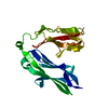
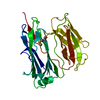
| Title | Crystal structure of the N-terminal Ig1-2 module of Human Receptor Protein Tyrosine Phosphatase Delta | ||||||
 Components Components | PTPRD PROTEIN | ||||||
 Keywords Keywords |  HYDROLASE HYDROLASE | ||||||
| Function / homology |  Function and homology information Function and homology informationtrans-synaptic signaling by trans-synaptic complex / cell surface receptor protein tyrosine phosphatase signaling pathway / Receptor-type tyrosine-protein phosphatases / presynaptic membrane assembly / synaptic membrane adhesion / regulation of postsynaptic density assembly / transmembrane receptor protein tyrosine phosphatase activity / presynapse assembly / Synaptic adhesion-like molecules / negative regulation of receptor signaling pathway via JAK-STAT ...trans-synaptic signaling by trans-synaptic complex / cell surface receptor protein tyrosine phosphatase signaling pathway / Receptor-type tyrosine-protein phosphatases / presynaptic membrane assembly / synaptic membrane adhesion / regulation of postsynaptic density assembly / transmembrane receptor protein tyrosine phosphatase activity / presynapse assembly / Synaptic adhesion-like molecules / negative regulation of receptor signaling pathway via JAK-STAT / phosphate-containing compound metabolic process / positive regulation of synapse assembly / positive regulation of dendrite morphogenesis / heterophilic cell-cell adhesion via plasma membrane cell adhesion molecules / regulation of immune response / cell adhesion molecule binding / hippocampal mossy fiber to CA3 synapse / protein dephosphorylation / protein-tyrosine-phosphatase / protein tyrosine phosphatase activity / Schaffer collateral - CA1 synapse / modulation of chemical synaptic transmission / neuron differentiation / presynaptic membrane / receptor complex / signaling receptor binding / glutamatergic synapse / extracellular exosome / plasma membraneSimilarity search - Function | ||||||
| Biological species |  HOMO SAPIENS (human) HOMO SAPIENS (human) | ||||||
| Method | X-RAY DIFFRACTION / SYNCHROTRON / MOLECULAR REPLACEMENT / Resolution: 1.98 Å | ||||||
 Authors Authors | Coles, C.H. / Shen, Y. / Tenney, A.P. / Siebold, C. / Sutton, G.C. / Lu, W. / Gallagher, J.T. / Jones, E.Y. / Flanagan, J.G. / Aricescu, A.R. | ||||||
 Citation Citation | Journal: Science / Year: 2011 Title: Proteoglycan-Specific Molecular Switch for Rptp Sigma Clustering and Neuronal Extension. Authors: Coles, C.H. / Shen, Y. / Tenney, A.P. / Siebold, C. / Sutton, G.C. / Lu, W. / Gallagher, J.T. / Jones, E.Y. / Flanagan, J.G. / Aricescu, A.R. | ||||||
| History |
|
- Structure visualization
| Structure viewer | Molecule: MolmilJmol/JSmol |
|---|
- Downloads & links
-Download
| PDBx/mmCIF format | 2yd7.cif.gz | 172 KB | Display | PDBx/mmCIF format |
|---|---|---|---|---|
| PDB format | pdb2yd7.ent.gz | 137.2 KB | Display | PDB format |
| PDBx/mmJSON format | 2yd7.json.gz | Tree view | PDBx/mmJSON format | |
| Others |  Other downloads Other downloads |
-Validation report
| Arichive directory | https://data.pdbj.org/pub/pdb/validation_reports/yd/2yd7ftp://data.pdbj.org/pub/pdb/validation_reports/yd/2yd7 | HTTPS FTP |
|---|
-Related structure data
| Related structure data | 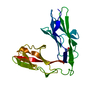 2yd1C  2yd2C  2yd3C  2yd4SC  2yd5C 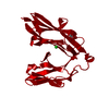 2yd6C  2yd8C  2yd9C C: citing same article ( S: Starting model for refinement |
|---|---|
| Similar structure data |


-Links
 PDBj
PDBj


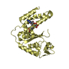
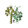
- Assembly
| Deposited unit | 
| ||||||||
|---|---|---|---|---|---|---|---|---|---|
| 1 | 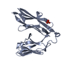
| ||||||||
| 2 | 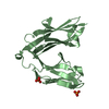
| ||||||||
| Unit cell |
| ||||||||
| Noncrystallographic symmetry (NCS) | NCS oper: (Code: given Matrix: (-0.9995, 0.0203, -0.026), Vector : |
-Components
| #1: Protein | Mass: 23480.199 Da / Num. of mol.: 2 / Fragment: IG1-2, RESIDUES 21-220 Source method: isolated from a genetically manipulated source Source: (gene. exp.) HOMO SAPIENS (human) / Cell line (production host): HEK293T / Production host: HOMO SAPIENS (human)References: UniProt: Q3KPJ2, UniProt: P23468*PLUS, protein-tyrosine-phosphatase#2: Chemical | Phosphate  Mass: 94.971 Da / Num. of mol.: 3 / Source method: obtained synthetically / Formula: PO4 Mass: 94.971 Da / Num. of mol.: 3 / Source method: obtained synthetically / Formula: PO4#3: Water | ChemComp-HOH / | Water Mass: 18.015 Da / Num. of mol.: 140 / Source method: isolated from a natural source / Formula: H2O Mass: 18.015 Da / Num. of mol.: 140 / Source method: isolated from a natural source / Formula: H2OSequence details | THE N-TERMINAL THREE AMINO ACID RESIDUES (ETG) AND THE C- TERMINAL NINE AMINO ACID RESIDUES ...THE N-TERMINAL THREE AMINO ACID RESIDUES (ETG) AND THE C- TERMINAL NINE AMINO ACID RESIDUES (GTKHHHHHH) DERIVE FROM THE PHLSEC VECTOR. | |
|---|
-Experimental details
-Experiment
| Experiment | Method: X-RAY DIFFRACTION / Number of used crystals: 1 |
|---|
- Sample preparation
| Crystal | Density Matthews: 2.33 Å3/Da / Density % sol: 48 % / Description: NONE |
|---|---|
| Crystal grow | Details: 20% W/V PEG 3350, 0.2M AMMONIUM DI-HYDROGEN PHOSPHATE . |
-Data collection
| Diffraction | Mean temperature: 100 K |
|---|---|
| Diffraction source | Source: SYNCHROTRON / Site: Diamond  / Beamline: I02 / Wavelength: 0.9795 / Beamline: I02 / Wavelength: 0.9795 |
| Detector | Type: ADSC CCD / Detector: CCD |
| Radiation | Protocol: SINGLE WAVELENGTH / Monochromatic (M) / Laue (L): M / Scattering type: x-ray |
| Radiation wavelength | Wavelength: 0.9795 Å / Relative weight: 1 |
| Reflection | Resolution: 2→50 Å / Num. obs: 30633 / % possible obs: 99.8 % / Observed criterion σ(I): 0 / Redundancy: 3.6 % / Biso Wilson estimate: 29.71 Å2 / Rmerge(I) obs: 0.07 / Net I/σ(I): 16.8 |
| Reflection shell | Resolution: 2→2.06 Å / Redundancy: 3.6 % / Rmerge(I) obs: 0.6 / Mean I/σ(I) obs: 2.1 / % possible all: 99.8 |
- Processing
| Software |
| ||||||||||||||||||||||||||||||||||||||||||||||||||||||||||||||||||||||||||||||||||||||||||||||||||||||||||||||||||||||||||||||||||||||||||||||||||||||||||||||||||||||||||||||||||||||
|---|---|---|---|---|---|---|---|---|---|---|---|---|---|---|---|---|---|---|---|---|---|---|---|---|---|---|---|---|---|---|---|---|---|---|---|---|---|---|---|---|---|---|---|---|---|---|---|---|---|---|---|---|---|---|---|---|---|---|---|---|---|---|---|---|---|---|---|---|---|---|---|---|---|---|---|---|---|---|---|---|---|---|---|---|---|---|---|---|---|---|---|---|---|---|---|---|---|---|---|---|---|---|---|---|---|---|---|---|---|---|---|---|---|---|---|---|---|---|---|---|---|---|---|---|---|---|---|---|---|---|---|---|---|---|---|---|---|---|---|---|---|---|---|---|---|---|---|---|---|---|---|---|---|---|---|---|---|---|---|---|---|---|---|---|---|---|---|---|---|---|---|---|---|---|---|---|---|---|---|---|---|---|---|
| Refinement | Method to determine structure: MOLECULAR REPLACEMENT Starting model: PDB ENTRY 2YD4 Resolution: 1.98→85.61 Å / Cor.coef. Fo:Fc: 0.952 / Cor.coef. Fo:Fc free: 0.937 / SU B: 9.417 / SU ML: 0.133 / Cross valid method: THROUGHOUT / ESU R: 0.198 / ESU R Free: 0.168 / Stereochemistry target values: MAXIMUM LIKELIHOOD Details: HYDROGENS HAVE BEEN ADDED IN THE RIDING POSITIONS. CLEAR ELECTRON DENSITY IS NOT VISIBLE FOR AMINO ACID RESIDUES 170-172 CHAIN A AND 169-173 CHAIN B. THESE RESIDUES ARE NOT INCLUDED IN THIS ...Details: HYDROGENS HAVE BEEN ADDED IN THE RIDING POSITIONS. CLEAR ELECTRON DENSITY IS NOT VISIBLE FOR AMINO ACID RESIDUES 170-172 CHAIN A AND 169-173 CHAIN B. THESE RESIDUES ARE NOT INCLUDED IN THIS CRYSTAL STRUCTURE. ARG 220 SIDECHAINS ARE DISORDERED AND THEREFORE NOT INCLUDED IN THE MODEL.
| ||||||||||||||||||||||||||||||||||||||||||||||||||||||||||||||||||||||||||||||||||||||||||||||||||||||||||||||||||||||||||||||||||||||||||||||||||||||||||||||||||||||||||||||||||||||
| Solvent computation | Ion probe radii: 0.8 Å / Shrinkage radii: 0.8 Å / VDW probe radii: 1.2 Å / Solvent model: MASK | ||||||||||||||||||||||||||||||||||||||||||||||||||||||||||||||||||||||||||||||||||||||||||||||||||||||||||||||||||||||||||||||||||||||||||||||||||||||||||||||||||||||||||||||||||||||
| Displacement parameters | Biso mean: 38.258 Å2
| ||||||||||||||||||||||||||||||||||||||||||||||||||||||||||||||||||||||||||||||||||||||||||||||||||||||||||||||||||||||||||||||||||||||||||||||||||||||||||||||||||||||||||||||||||||||
| Refinement step | Cycle: LAST / Resolution: 1.98→85.61 Å
| ||||||||||||||||||||||||||||||||||||||||||||||||||||||||||||||||||||||||||||||||||||||||||||||||||||||||||||||||||||||||||||||||||||||||||||||||||||||||||||||||||||||||||||||||||||||
| Refine LS restraints |
|