 Movie
Movie Controller

[English] 日本語
 Yorodumi
- PDB-5hlo: Crystal structure of calcium and zinc-bound human S100A8 in space... -
+ Open data

- Basic information
| Entry | Database: PDB / ID: 5hlo | ||||||
|---|---|---|---|---|---|---|---|
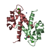
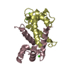
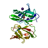
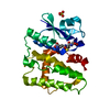
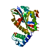
| Title | Crystal structure of calcium and zinc-bound human S100A8 in space group C2221 | ||||||
 Components Components | Protein S100-A8 | ||||||
 Keywords Keywords |  SIGNALING PROTEIN / S100 protein / calcium / zinc / oligomer SIGNALING PROTEIN / S100 protein / calcium / zinc / oligomer | ||||||
| Function / homology |  Function and homology information Function and homology informationsequestering of zinc ion / calprotectin complex / neutrophil aggregation / positive regulation of peptide secretion / autocrine signaling / Toll-like receptor 4 binding / chronic inflammatory response / Metal sequestration by antimicrobial proteins / leukocyte migration involved in inflammatory response / peptide secretion ...sequestering of zinc ion / calprotectin complex / neutrophil aggregation / positive regulation of peptide secretion / autocrine signaling / Toll-like receptor 4 binding / chronic inflammatory response / Metal sequestration by antimicrobial proteins / leukocyte migration involved in inflammatory response / peptide secretion / RAGE receptor binding / Regulation of TLR by endogenous ligand / astrocyte development / MyD88 deficiency (TLR2/4) / arachidonic acid binding / intermediate filament cytoskeleton / IRAK4 deficiency (TLR2/4) / MyD88:MAL(TIRAP) cascade initiated on plasma membrane / response to zinc ion / regulation of toll-like receptor signaling pathway / regulation of cytoskeleton organization / peptidyl-cysteine S-nitrosylation / RHO GTPases Activate NADPH Oxidases / defense response to fungus / positive regulation of intrinsic apoptotic signaling pathway / neutrophil chemotaxis / autophagy / positive regulation of inflammatory response / calcium-dependent protein binding / activation of cysteine-type endopeptidase activity involved in apoptotic process / positive regulation of NF-kappaB transcription factor activity / ER-Phagosome pathway / positive regulation of cell growth / microtubule binding / response to ethanol / collagen-containing extracellular matrix / secretory granule lumen / response to lipopolysaccharide / cytoskeleton / defense response to bacterium / inflammatory response / innate immune response / apoptotic process / calcium ion binding / Neutrophil degranulation / extracellular space / extracellular exosome / zinc ion binding / extracellular region / nucleus / plasma membrane / cytosolSimilarity search - Function | ||||||
| Biological species |  Homo sapiens (human) Homo sapiens (human) | ||||||
| Method | X-RAY DIFFRACTION / SYNCHROTRON / MOLECULAR REPLACEMENT / Resolution: 2.1 Å | ||||||
 Authors Authors | Lin, H. / Andersen, G.R. / Yatime, L. | ||||||
 Citation Citation | Journal: Bmc Struct.Biol. / Year: 2016 Title: Crystal structure of human S100A8 in complex with zinc and calcium. Authors: Lin, H. / Andersen, G.R. / Yatime, L. | ||||||
| History |
|
- Structure visualization
| Structure viewer | Molecule: MolmilJmol/JSmol |
|---|
- Downloads & links
-Download
| PDBx/mmCIF format | 5hlo.cif.gz | 173 KB | Display | PDBx/mmCIF format |
|---|---|---|---|---|
| PDB format | pdb5hlo.ent.gz | 137.7 KB | Display | PDB format |
| PDBx/mmJSON format | 5hlo.json.gz | Tree view | PDBx/mmJSON format | |
| Others |  Other downloads Other downloads |
-Validation report
| Arichive directory | https://data.pdbj.org/pub/pdb/validation_reports/hl/5hloftp://data.pdbj.org/pub/pdb/validation_reports/hl/5hlo | HTTPS FTP |
|---|
-Related structure data
| Related structure data | 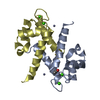 5hlvC 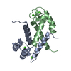 1mr8S S: Starting model for refinement C: citing same article ( |
|---|---|
| Similar structure data |


-Links
 PDBj
PDBj


- Assembly
| Deposited unit | 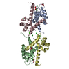
| ||||||||
|---|---|---|---|---|---|---|---|---|---|
| 1 | 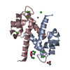
| ||||||||
| 2 | 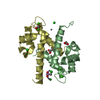
| ||||||||
| Unit cell |
| ||||||||
| Components on special symmetry positions |
|
-Components
-Protein , 1 types, 4 molecules ABCD
| #1: Protein | Mass: 10924.606 Da / Num. of mol.: 4 Source method: isolated from a genetically manipulated source Source: (gene. exp.) Homo sapiens (human) / Gene: S100A8, CAGA, CFAG, MRP8 / Plasmid: pETM13 / Production host:  Escherichia coli BL21(DE3) (bacteria) / References: UniProt: P05109 Escherichia coli BL21(DE3) (bacteria) / References: UniProt: P05109 |
|---|
-Non-polymers , 6 types, 223 molecules 


| #2: Chemical | ChemComp-CA /  Mass: 40.078 Da / Num. of mol.: 8 / Source method: obtained synthetically / Formula: Ca Mass: 40.078 Da / Num. of mol.: 8 / Source method: obtained synthetically / Formula: Ca#3: Chemical | ChemComp-ZN /  Mass: 65.409 Da / Num. of mol.: 8 / Source method: obtained synthetically / Formula: Zn Mass: 65.409 Da / Num. of mol.: 8 / Source method: obtained synthetically / Formula: Zn#4: Chemical | Cacodylic acid Mass: 136.989 Da / Num. of mol.: 2 / Source method: obtained synthetically / Formula: C2H6AsO2 Mass: 136.989 Da / Num. of mol.: 2 / Source method: obtained synthetically / Formula: C2H6AsO2#5: Chemical | ChemComp-CL / Chloride Mass: 35.453 Da / Num. of mol.: 8 / Source method: obtained synthetically / Formula: Cl Mass: 35.453 Da / Num. of mol.: 8 / Source method: obtained synthetically / Formula: Cl#6: Chemical | ChemComp-ACT / Acetate Mass: 59.044 Da / Num. of mol.: 4 / Source method: obtained synthetically / Formula: C2H3O2 Mass: 59.044 Da / Num. of mol.: 4 / Source method: obtained synthetically / Formula: C2H3O2#7: Water | ChemComp-HOH / | WaterMass: 18.015 Da / Num. of mol.: 193 / Source method: isolated from a natural source / Formula: H2O |
|---|
-Experimental details
-Experiment
| Experiment | Method: X-RAY DIFFRACTION |
|---|
- Sample preparation
| Crystal | Density Matthews: 2.87 Å3/Da / Density % sol: 57.2 % |
|---|---|
| Crystal grow | Temperature: 293 K / Method: vapor diffusion, sitting drop / pH: 6.5 Details: 5 mM ZnCl2, 0.1 M Na cacodylate pH 6.5, 8% PEG 8000 |
-Data collection
| Diffraction | Mean temperature: 100 K |
|---|---|
| Diffraction source | Source: SYNCHROTRON / Site: MAX II  / Beamline: I911-3 / Wavelength: 1 Å / Beamline: I911-3 / Wavelength: 1 Å |
| Detector | Type: MARMOSAIC 225 mm CCD / Detector: CCD / Date: Jun 29, 2014 |
| Radiation | Protocol: SINGLE WAVELENGTH / Monochromatic (M) / Laue (L): M / Scattering type: x-ray |
| Radiation wavelength | Wavelength: 1 Å / Relative weight: 1 |
| Reflection | Resolution: 2.1→50 Å / Num. obs: 29069 / % possible obs: 98.5 % / Redundancy: 8 % / Rsym value: 0.095 / Net I/σ(I): 16.47 |
| Reflection shell | Resolution: 2.1→2.2 Å / Redundancy: 8.2 % / Mean I/σ(I) obs: 3.04 / % possible all: 97.8 |
- Processing
| Software |
| ||||||||||||||||||||||||||||||||||||||||||||||||||||||||||||||||||||||||||||||||||||||||||||||||||||||||||||||||||||||||||||||||||
|---|---|---|---|---|---|---|---|---|---|---|---|---|---|---|---|---|---|---|---|---|---|---|---|---|---|---|---|---|---|---|---|---|---|---|---|---|---|---|---|---|---|---|---|---|---|---|---|---|---|---|---|---|---|---|---|---|---|---|---|---|---|---|---|---|---|---|---|---|---|---|---|---|---|---|---|---|---|---|---|---|---|---|---|---|---|---|---|---|---|---|---|---|---|---|---|---|---|---|---|---|---|---|---|---|---|---|---|---|---|---|---|---|---|---|---|---|---|---|---|---|---|---|---|---|---|---|---|---|---|---|---|
| Refinement | Method to determine structure: MOLECULAR REPLACEMENT Starting model: 1MR8 Resolution: 2.1→49.2 Å / SU ML: 0.25 / Cross valid method: FREE R-VALUE / σ(F): 2 / Phase error: 21.03 / Stereochemistry target values: ML
| ||||||||||||||||||||||||||||||||||||||||||||||||||||||||||||||||||||||||||||||||||||||||||||||||||||||||||||||||||||||||||||||||||
| Solvent computation | Shrinkage radii: 0.9 Å / VDW probe radii: 1.11 Å / Solvent model: FLAT BULK SOLVENT MODEL | ||||||||||||||||||||||||||||||||||||||||||||||||||||||||||||||||||||||||||||||||||||||||||||||||||||||||||||||||||||||||||||||||||
| Refinement step | Cycle: LAST / Resolution: 2.1→49.2 Å
| ||||||||||||||||||||||||||||||||||||||||||||||||||||||||||||||||||||||||||||||||||||||||||||||||||||||||||||||||||||||||||||||||||
| Refine LS restraints |
| ||||||||||||||||||||||||||||||||||||||||||||||||||||||||||||||||||||||||||||||||||||||||||||||||||||||||||||||||||||||||||||||||||
| LS refinement shell |
| ||||||||||||||||||||||||||||||||||||||||||||||||||||||||||||||||||||||||||||||||||||||||||||||||||||||||||||||||||||||||||||||||||
| Refinement TLS params. | Method: refined / Refine-ID: X-RAY DIFFRACTION
| ||||||||||||||||||||||||||||||||||||||||||||||||||||||||||||||||||||||||||||||||||||||||||||||||||||||||||||||||||||||||||||||||||
| Refinement TLS group |
|
