Loss of function of TP53 in cancer due to loss of tetramerization ability / Regulation of TP53 Expression / signal transduction by p53 class mediator / negative regulation of G1 to G0 transition / negative regulation of glucose catabolic process to lactate via pyruvate / Transcriptional activation of cell cycle inhibitor p21 / regulation of intrinsic apoptotic signaling pathway by p53 class mediator / Activation of NOXA and translocation to mitochondria / negative regulation of pentose-phosphate shunt / ATP-dependent DNA/DNA annealing activity ...Loss of function of TP53 in cancer due to loss of tetramerization ability / Regulation of TP53 Expression / signal transduction by p53 class mediator / negative regulation of G1 to G0 transition / negative regulation of glucose catabolic process to lactate via pyruvate / Transcriptional activation of cell cycle inhibitor p21 / regulation of intrinsic apoptotic signaling pathway by p53 class mediator / Activation of NOXA and translocation to mitochondria / negative regulation of pentose-phosphate shunt / ATP-dependent DNA/DNA annealing activity / negative regulation of helicase activity / regulation of cell cycle G2/M phase transition / intrinsic apoptotic signaling pathway in response to hypoxia / regulation of fibroblast apoptotic process / oxidative stress-induced premature senescence / oligodendrocyte apoptotic process / negative regulation of miRNA processing / positive regulation of thymocyte apoptotic process / glucose catabolic process to lactate via pyruvate / regulation of tissue remodeling / positive regulation of mitochondrial membrane permeability / negative regulation of mitophagy / positive regulation of programmed necrotic cell death / mRNA transcription / bone marrow development / circadian behavior / histone deacetylase regulator activity / regulation of mitochondrial membrane permeability involved in apoptotic process / RUNX3 regulates CDKN1A transcription / germ cell nucleus / regulation of DNA damage response, signal transduction by p53 class mediator / TP53 regulates transcription of additional cell cycle genes whose exact role in the p53 pathway remain uncertain / TP53 Regulates Transcription of Death Receptors and Ligands / Activation of PUMA and translocation to mitochondria / DNA damage response, signal transduction by p53 class mediator resulting in transcription of p21 class mediator / negative regulation of glial cell proliferation / Formation of Senescence-Associated Heterochromatin Foci (SAHF) / negative regulation of neuroblast proliferation / Regulation of TP53 Activity through Association with Co-factors / mitochondrial DNA repair / T cell lineage commitment / positive regulation of execution phase of apoptosis / negative regulation of DNA replication / ER overload response / B cell lineage commitment / thymocyte apoptotic process / TP53 regulates transcription of several additional cell death genes whose specific roles in p53-dependent apoptosis remain uncertain / positive regulation of cardiac muscle cell apoptotic process / TP53 Regulates Transcription of Caspase Activators and Caspases / T cell proliferation involved in immune response / cardiac septum morphogenesis / entrainment of circadian clock by photoperiod / PI5P Regulates TP53 Acetylation / Association of TriC/CCT with target proteins during biosynthesis / Zygotic genome activation (ZGA) / necroptotic process / rRNA transcription / positive regulation of release of cytochrome c from mitochondria / TP53 Regulates Transcription of Genes Involved in Cytochrome C Release / TFIID-class transcription factor complex binding / mitophagy / SUMOylation of transcription factors / negative regulation of telomere maintenance via telomerase / general transcription initiation factor binding / intrinsic apoptotic signaling pathway by p53 class mediator / Transcriptional Regulation by VENTX / response to X-ray / DNA damage response, signal transduction by p53 class mediator / replicative senescence / chromosome organization / neuroblast proliferation / intrinsic apoptotic signaling pathway in response to endoplasmic reticulum stress / cellular response to UV-C / : / hematopoietic stem cell differentiation / negative regulation of reactive oxygen species metabolic process / intrinsic apoptotic signaling pathway in response to DNA damage by p53 class mediator / glial cell proliferation / embryonic organ development / positive regulation of RNA polymerase II transcription preinitiation complex assembly / Pyroptosis / cis-regulatory region sequence-specific DNA binding / hematopoietic progenitor cell differentiation / cellular response to glucose starvation / cellular response to actinomycin D / TP53 Regulates Transcription of Genes Involved in G1 Cell Cycle Arrest / somitogenesis / type II interferon-mediated signaling pathway / positive regulation of intrinsic apoptotic signaling pathway / negative regulation of stem cell proliferation / core promoter sequence-specific DNA binding / gastrulation / negative regulation of fibroblast proliferation / MDM2/MDM4 family protein binding / cardiac muscle cell apoptotic process / transcription initiation-coupled chromatin remodeling / 14-3-3 protein binding / mitotic G1 DNA damage checkpoint signaling / Regulation of TP53 Activity through Acetylation / response to salt stress Similarity search - Function
Mass: 18.015 Da / Num. of mol.: 644 / Source method: isolated from a natural source / Formula: H2O
-
Experimental details
-
Experiment
Experiment
Method: X-RAY DIFFRACTION / Number of used crystals: 1
-
Sample preparation
Crystal
Density Matthews: 2.2 Å3/Da / Density % sol: 45 % / Description: NONE
Crystal grow
Details: PROTEIN SOLUTION: 5 MG/ML PROTEIN IN 20 MM CITRATE BUFFER PH 6.1, 150 MM NACL, 10 MM DTT. CRYSTALLIZATION BUFFER: 26% (W/V) PEG 3350, 43 MM SODIUM ACETATE AND 100 MM HEPES, PH 7.5
Method to determine structure: MOLECULAR REPLACEMENT / Resolution: 1.9→42.212 Å / σ(F): 1.35 / Phase error: 34.69 / Stereochemistry target values: TWIN_LSQ_F Details: IN THE UNBOUND P53 DNA-BINDING DOMAIN, THE LYS120 SIDE CHAIN IS HIGHLY FLEXIBLE. SIMILARLY, THE SIDE CHAIN OF ACETYLATED LYS120 SHOWED A HIGH DEGREE OF FLEXIBILITY, AND THE LACK OF DEFINED ...Details: IN THE UNBOUND P53 DNA-BINDING DOMAIN, THE LYS120 SIDE CHAIN IS HIGHLY FLEXIBLE. SIMILARLY, THE SIDE CHAIN OF ACETYLATED LYS120 SHOWED A HIGH DEGREE OF FLEXIBILITY, AND THE LACK OF DEFINED ELECTRON DENSITY PREVENTED UNAMBIGUOUS MODELING OF THE ACETYLATED SIDE CHAIN.
Rfactor
Num. reflection
% reflection
Rfree
0.226
6156
6 %
Rwork
0.1744
-
-
obs
0.1768
101869
83.31 %
Solvent computation
Shrinkage radii: 0.9 Å / VDW probe radii: 1.11 Å / Solvent model: FLAT BULK SOLVENT MODEL / Bsol: 29.326 Å2 / ksol: 0.343 e/Å3
Displacement parameters
Baniso -1
Baniso -2
Baniso -3
1-
-6.1068 Å2
0 Å2
-2.0096 Å2
2-
-
11.937 Å2
0 Å2
3-
-
-
-5.8302 Å2
Refinement step
Cycle: LAST / Resolution: 1.9→42.212 Å
Protein
Nucleic acid
Ligand
Solvent
Total
Num. atoms
6056
0
4
644
6704
Refine LS restraints
Refine-ID
Type
Dev ideal
Number
X-RAY DIFFRACTION
f_bond_d
0.006
6221
X-RAY DIFFRACTION
f_angle_d
0.97
8451
X-RAY DIFFRACTION
f_dihedral_angle_d
15.91
2327
X-RAY DIFFRACTION
f_chiral_restr
0.064
924
X-RAY DIFFRACTION
f_plane_restr
0.004
1122
LS refinement shell
Resolution (Å)
Rfactor Rfree
Num. reflection Rfree
Rfactor Rwork
Num. reflection Rwork
Refine-ID
% reflection obs (%)
1.9001-1.9328
0.3184
220
0.2607
4823
X-RAY DIFFRACTION
79
1.9328-1.968
0.2798
259
0.2569
4771
X-RAY DIFFRACTION
78
1.968-2.0058
0.2969
254
0.2439
4824
X-RAY DIFFRACTION
79
2.0058-2.0467
0.2886
267
0.231
4864
X-RAY DIFFRACTION
78
2.0467-2.0912
0.2581
288
0.2205
4725
X-RAY DIFFRACTION
78
2.0912-2.1399
0.2656
274
0.2177
4790
X-RAY DIFFRACTION
79
2.1399-2.1934
0.2617
255
0.2098
4804
X-RAY DIFFRACTION
79
2.1934-2.2527
0.2371
227
0.2073
4896
X-RAY DIFFRACTION
79
2.2527-2.3189
0.2594
230
0.2038
4910
X-RAY DIFFRACTION
80
2.3189-2.3938
0.2682
238
0.1925
4826
X-RAY DIFFRACTION
80
2.3938-2.4793
0.2362
275
0.1973
4848
X-RAY DIFFRACTION
79
2.4793-2.5785
0.2736
243
0.1938
4914
X-RAY DIFFRACTION
80
2.5785-2.6958
0.231
244
0.1859
4809
X-RAY DIFFRACTION
80
2.6958-2.8379
0.2489
288
0.1829
4828
X-RAY DIFFRACTION
79
2.8379-3.0155
0.2015
213
0.1663
4931
X-RAY DIFFRACTION
80
3.0155-3.2482
0.2119
268
0.157
4848
X-RAY DIFFRACTION
79
3.2482-3.5746
0.2065
250
0.1342
4855
X-RAY DIFFRACTION
80
3.5746-4.091
0.1707
282
0.1279
4787
X-RAY DIFFRACTION
78
4.091-5.1508
0.1776
229
0.1197
4566
X-RAY DIFFRACTION
75
5.1508-32.1185
0.2038
296
0.1542
5138
X-RAY DIFFRACTION
84
+
About Yorodumi
-
News
-
Feb 9, 2022. New format data for meta-information of EMDB entries
New format data for meta-information of EMDB entries
Version 3 of the EMDB header file is now the official format.
The previous official version 1.9 will be removed from the archive.
In the structure databanks used in Yorodumi, some data are registered as the other names, "COVID-19 virus" and "2019-nCoV". Here are the details of the virus and the list of structure data.
Jan 31, 2019. EMDB accession codes are about to change! (news from PDBe EMDB page)
EMDB accession codes are about to change! (news from PDBe EMDB page)
The allocation of 4 digits for EMDB accession codes will soon come to an end. Whilst these codes will remain in use, new EMDB accession codes will include an additional digit and will expand incrementally as the available range of codes is exhausted. The current 4-digit format prefixed with “EMD-” (i.e. EMD-XXXX) will advance to a 5-digit format (i.e. EMD-XXXXX), and so on. It is currently estimated that the 4-digit codes will be depleted around Spring 2019, at which point the 5-digit format will come into force.
The EM Navigator/Yorodumi systems omit the EMD- prefix.
Related info.:Q: What is EMD? / ID/Accession-code notation in Yorodumi/EM Navigator
Yorodumi is a browser for structure data from EMDB, PDB, SASBDB, etc.
This page is also the successor to EM Navigator detail page, and also detail information page/front-end page for Omokage search.
The word "yorodu" (or yorozu) is an old Japanese word meaning "ten thousand". "mi" (miru) is to see.
Related info.:EMDB / PDB / SASBDB / Comparison of 3 databanks / Yorodumi Search / Aug 31, 2016. New EM Navigator & Yorodumi / Yorodumi Papers / Jmol/JSmol / Function and homology information / Changes in new EM Navigator and Yorodumi
 Movie
Movie Controller


 Open data
 Basic information
 Components
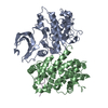
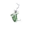
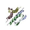
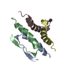
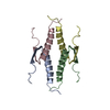
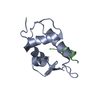
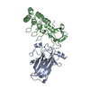
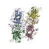
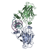
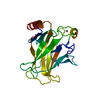
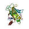
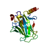
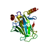
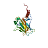
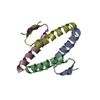
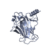
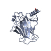
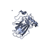
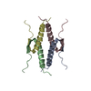
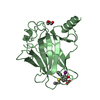
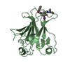
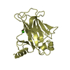
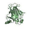
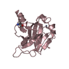
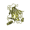
 P53
P53  Keywords
 Function and homology information
 Authors
 Citation
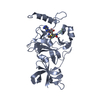
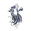
 Structure visualization
 Downloads & links
 Other downloads
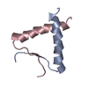


 PDBj
PDBj


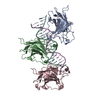
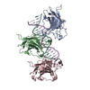
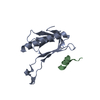
 Assembly


 Mass: 65.409 Da / Num. of mol.: 4 / Source method: obtained synthetically / Formula: Zn
 Mass: 18.015 Da / Num. of mol.: 644 / Source method: isolated from a natural source / Formula: H2O
 Sample preparation
 / Beamline: ID14-2 / Wavelength: 0.979
  Processing