 Movie
Movie Controller

[English] 日本語
 Yorodumi
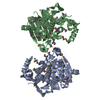
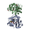
- PDB-2xyl: CELLULOMONAS FIMI XYLANASE/CELLULASE COMPLEXED WITH 2-DEOXY-2-FLU... -
+ Open data

- Basic information
| Entry | Database: PDB / ID: 2xyl | |||||||||
|---|---|---|---|---|---|---|---|---|---|---|
| Title | CELLULOMONAS FIMI XYLANASE/CELLULASE COMPLEXED WITH 2-DEOXY-2-FLUORO-XYLOBIOSE | |||||||||
 Components Components | BETA-1,4-GLYCANASE | |||||||||
 Keywords Keywords |  HYDROLASE / O-GLYCOSYL / XYLANASE/CELLULASE / A/B BARREL / CELLULOSE DEGRADATION HYDROLASE / O-GLYCOSYL / XYLANASE/CELLULASE / A/B BARREL / CELLULOSE DEGRADATION | |||||||||
| Function / homology |  Function and homology information Function and homology informationcellulose 1,4-beta-cellobiosidase (non-reducing end) / cellulose 1,4-beta-cellobiosidase activity / polysaccharide binding / endo-1,4-beta-xylanase activity / endo-1,4-beta-xylanase / xylan catabolic process / cellulose catabolic processSimilarity search - Function | |||||||||
| Biological species |  Cellulomonas fimi (bacteria) Cellulomonas fimi (bacteria) | |||||||||
| Method | X-RAY DIFFRACTION / MOLECULAR SUBSTITUTION / Resolution: 1.9 Å | |||||||||
 Authors Authors | Monem, V. / Birsan, C. / Warren, R.A.J. / Withers, S.G. / Rose, D.R. | |||||||||
 Citation Citation | Journal: Biochemistry / Year: 1998 Title: Exploring the cellulose/xylan specificity of the beta-1,4-glycanase cex from Cellulomonas fimi through crystallography and mutation. Authors: Notenboom, V. / Birsan, C. / Warren, R.A. / Withers, S.G. / Rose, D.R. | |||||||||
| History |
|
- Structure visualization
| Structure viewer | Molecule: MolmilJmol/JSmol |
|---|
- Downloads & links
-Download
| PDBx/mmCIF format | 2xyl.cif.gz | 71.4 KB | Display | PDBx/mmCIF format |
|---|---|---|---|---|
| PDB format | pdb2xyl.ent.gz | 55.2 KB | Display | PDB format |
| PDBx/mmJSON format | 2xyl.json.gz | Tree view | PDBx/mmJSON format | |
| Others |  Other downloads Other downloads |
-Validation report
| Arichive directory | https://data.pdbj.org/pub/pdb/validation_reports/xy/2xylftp://data.pdbj.org/pub/pdb/validation_reports/xy/2xyl | HTTPS FTP |
|---|
-Related structure data
| Related structure data | 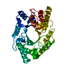 2exoS S: Starting model for refinement |
|---|---|
| Similar structure data |


-Links
 PDBj
PDBj

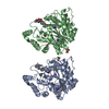
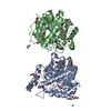
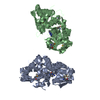
- Assembly
| Deposited unit | 
| ||||||||
|---|---|---|---|---|---|---|---|---|---|
| 1 | 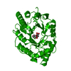
| ||||||||
| Unit cell |
|
-Components
| #1: Protein | Mass: 34051.941 Da / Num. of mol.: 1 / Fragment: CATALYTIC DOMAIN Source method: isolated from a genetically manipulated source Source: (gene. exp.) Cellulomonas fimi (bacteria) / Production host: Escherichia coli (E. coli)References: GenBank: 144429, UniProt: P07986*PLUS, cellulose 1,4-beta-cellobiosidase (non-reducing end) |
|---|---|
| #2: Polysaccharide | beta-D-xylopyranose-(1-4)-2-deoxy-2-fluoro-alpha-D-xylopyranose / Mass: 284.236 Da / Num. of mol.: 1 Source method: isolated from a genetically manipulated source |
| #3: Water | ChemComp-HOH / Water Mass: 18.015 Da / Num. of mol.: 122 / Source method: isolated from a natural source / Formula: H2O Mass: 18.015 Da / Num. of mol.: 122 / Source method: isolated from a natural source / Formula: H2O |
-Experimental details
-Experiment
| Experiment | Method: X-RAY DIFFRACTION / Number of used crystals: 1 |
|---|
- Sample preparation
| Crystal | Density Matthews: 2.32 Å3/Da / Density % sol: 46.97 % | ||||||||||||||||||||||||||||||||||||
|---|---|---|---|---|---|---|---|---|---|---|---|---|---|---|---|---|---|---|---|---|---|---|---|---|---|---|---|---|---|---|---|---|---|---|---|---|---|
| Crystal grow | pH: 4.6 Details: 7-10% PEG 4K, .1M NAACETATE PH4.6 60 MG/ML PROTEIN CONCENTRATION SOAK 48 HRS WITH 2F-XYLOBIOSE-DNP | ||||||||||||||||||||||||||||||||||||
| Crystal grow | *PLUS Method: vapor diffusion, hanging drop / Details: Bedarkar, S., (1992) J.Mol. Biol, 228, 693. | ||||||||||||||||||||||||||||||||||||
| Components of the solutions | *PLUS
|
-Data collection
| Diffraction | Mean temperature: 298 K |
|---|---|
| Diffraction source | Source: ROTATING ANODE / Type: RIGAKU RUH2R / Wavelength: 1.5418 |
| Detector | Type: XUONG-HAMLIN MULTIWIRE / Detector: AREA DETECTOR / Date: Apr 1, 1996 |
| Radiation | Monochromatic (M) / Laue (L): M / Scattering type: x-ray |
| Radiation wavelength | Wavelength: 1.5418 Å / Relative weight: 1 |
| Reflection | Highest resolution: 1.9 Å / Num. obs: 25857 / % possible obs: 99.94 % / Observed criterion σ(I): 2 / Redundancy: 7.64 % / Rsym value: 0.0803 / Net I/σ(I): 16.19 |
| Reflection shell | Resolution: 1.9→2.05 Å / Redundancy: 5.06 % / Mean I/σ(I) obs: 3.58 / Rsym value: 0.2703 / % possible all: 99.9 |
| Reflection | *PLUS Num. measured all: 197626 / Rmerge(I) obs: 0.0803 |
| Reflection shell | *PLUS Highest resolution: 1.9 Å / % possible obs: 99.9 % / Num. unique obs: 5050 / Num. measured obs: 25539 / Rmerge(I) obs: 0.2703 |
- Processing
| Software |
| ||||||||||||||||||||||||||||||||||||||||||||||||||||||||||||
|---|---|---|---|---|---|---|---|---|---|---|---|---|---|---|---|---|---|---|---|---|---|---|---|---|---|---|---|---|---|---|---|---|---|---|---|---|---|---|---|---|---|---|---|---|---|---|---|---|---|---|---|---|---|---|---|---|---|---|---|---|---|
| Refinement | Method to determine structure: MOLECULAR SUBSTITUTION Starting model: PDB ENTRY 2EXO Resolution: 1.9→10 Å / Rfactor Rfree error: 0.006 / Data cutoff high absF: 1000000 / Data cutoff low absF: 0.0001 / Cross valid method: THROUGHOUT / σ(F): 2
| ||||||||||||||||||||||||||||||||||||||||||||||||||||||||||||
| Displacement parameters | Biso mean: 13 Å2 | ||||||||||||||||||||||||||||||||||||||||||||||||||||||||||||
| Refinement step | Cycle: LAST / Resolution: 1.9→10 Å
| ||||||||||||||||||||||||||||||||||||||||||||||||||||||||||||
| Refine LS restraints |
| ||||||||||||||||||||||||||||||||||||||||||||||||||||||||||||
| LS refinement shell | Resolution: 1.9→1.99 Å / Rfactor Rfree error: 0.023 / Total num. of bins used: 8
| ||||||||||||||||||||||||||||||||||||||||||||||||||||||||||||
| Xplor file |
| ||||||||||||||||||||||||||||||||||||||||||||||||||||||||||||
| Software | *PLUS Name: X-PLOR / Version: 3.851 / Classification: refinement | ||||||||||||||||||||||||||||||||||||||||||||||||||||||||||||
| Refinement | *PLUS Rfactor Rfree: 0.26 | ||||||||||||||||||||||||||||||||||||||||||||||||||||||||||||
| Solvent computation | *PLUS | ||||||||||||||||||||||||||||||||||||||||||||||||||||||||||||
| Displacement parameters | *PLUS | ||||||||||||||||||||||||||||||||||||||||||||||||||||||||||||
| Refine LS restraints | *PLUS
|
