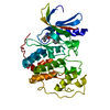
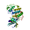
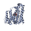
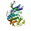
[English] 日本語
Yorodumi
- PDB-2xwv: SiaP complex -

+
Open data


ID or keywords:

Loading...

-
Basic information

Entry
Database: PDB / ID: 2xwv
TitleSiaP complex
ComponentsSIALIC ACID-BINDING PERIPLASMIC PROTEIN SIAP
KeywordsTRANSPORT PROTEIN / TRAP / SUGAR TRANSPORT
Function / homology
Function and homology information


: / transmembrane transport / outer membrane-bounded periplasmic space
Similarity search - Function
TRAP transporter solute receptor, DctP family / Bacterial extracellular solute-binding protein, family 7 / TRAP transporter solute receptor DctP / TRAP transporter solute receptor DctP superfamily / Bacterial extracellular solute-binding protein, family 7 / D-Maltodextrin-Binding Protein; domain 2 / 3-Layer(aba) Sandwich / Alpha Beta
Similarity search - Domain/homology
N-acetyl-beta-neuraminic acid / Sialic acid-binding periplasmic protein SiaP
Similarity search - Component
Biological speciesHAEMOPHILUS INFLUENZAE (bacteria)
MethodX-RAY DIFFRACTION / SYNCHROTRON / OTHER / Resolution: 1.05 Å
AuthorsFischer, M. / Hubbard, R.E.
CitationJournal: J.Biol.Chem. / Year: 2015
Title: Tripartite ATP-Independent Periplasmic (Trap) Transporters Use an Arginine-Mediated Selectivity Filter for High Affinity Substrate Binding.
Authors: Fischer, M. / Hopkins, A.P. / Severi, E. / Hawkhead, J. / Bawdon, D. / Watts, A.G. / Hubbard, R.E. / Thomas, G.H.
History
DepositionNov 5, 2010Deposition site: PDBE / Processing site: PDBE
Revision 1.0Nov 16, 2011Provider: repository / Type: Initial release
Revision 1.1Nov 23, 2011Group: Structure summary
Revision 1.2Sep 16, 2015Group: Database references
Revision 1.3Sep 23, 2015Group: Database references
Revision 1.4Nov 18, 2015Group: Database references
Revision 1.5Mar 6, 2019Group: Data collection / Experimental preparation / Other
Category: exptl_crystal_grow / pdbx_database_proc / pdbx_database_status
Item: _exptl_crystal_grow.temp / _pdbx_database_status.recvd_author_approval
Revision 1.6Jul 29, 2020Group: Data collection / Derived calculations ...Data collection / Derived calculations / Other / Structure summary
Category: chem_comp / entity ...chem_comp / entity / pdbx_chem_comp_identifier / pdbx_database_status / pdbx_entity_nonpoly / struct_site / struct_site_gen
Item: _chem_comp.name / _chem_comp.type ..._chem_comp.name / _chem_comp.type / _entity.pdbx_description / _pdbx_database_status.status_code_sf / _pdbx_entity_nonpoly.name
Description: Carbohydrate remediation / Provider: repository / Type: Remediation
Revision 1.7May 8, 2024Group: Data collection / Database references / Structure summary
Category: chem_comp / chem_comp_atom ...chem_comp / chem_comp_atom / chem_comp_bond / database_2
Item: _chem_comp.pdbx_synonyms / _database_2.pdbx_DOI / _database_2.pdbx_database_accession

-
Structure visualization

Structure viewerMolecule:
MolmilJmol/JSmol

Downloads & links

-
Assembly

Deposited unit
A: SIALIC ACID-BINDING PERIPLASMIC PROTEIN SIAP
hetero molecules


Theoretical massNumber of molelcules
Total (without water)35,3422
Polymers35,0331
Non-polymers3091
Water9,512528
1


  • Idetical with deposited unit
  • defined by author&software
TypeNameSymmetry operationNumber
identity operation1_555x,y,z1
MethodPISA
Unit cell
Length a, b, c (Å)47.723, 74.510, 86.930
Angle α, β, γ (deg.)90.00, 90.00, 90.00
Int Tables number19
Space group name H-MP212121

-
Components

#1: Protein SIALIC ACID-BINDING PERIPLASMIC PROTEIN SIAP / EXTRACYTOPLASMIC SOLUTE RECEPTOR PROTEIN SIAP / N-ACETYLNEURAMINIC-BINDING PROTEIN / NEU5AC-BINDING ...EXTRACYTOPLASMIC SOLUTE RECEPTOR PROTEIN SIAP / N-ACETYLNEURAMINIC-BINDING PROTEIN / NEU5AC-BINDING PROTEIN / SUBSTRATE BINDING PROTEIN


Mass: 35032.617 Da / Num. of mol.: 1
Source method: isolated from a genetically manipulated source
Source: (gene. exp.) HAEMOPHILUS INFLUENZAE (bacteria) / Production host: ESCHERICHIA COLI (E. coli) / Strain (production host): BL21(DE3) / Variant (production host): PLYSS PAH16 / References: UniProt: P44542
#2: Sugar ChemComp-SLB / N-acetyl-beta-neuraminic acid / N-acetylneuraminic acid / sialic acid / O-sialic acid / 5-N-ACETYL-BETA-D-NEURAMINIC ACID / BETA-SIALIC ACID / N-Acetylneuraminic acid


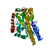
Type: D-saccharide, beta linking / Mass: 309.270 Da / Num. of mol.: 1
Source method: isolated from a genetically manipulated source
Formula: C11H19NO9
IdentifierTypeProgram
DNeup5AcbCONDENSED IUPAC CARBOHYDRATE SYMBOLGMML 1.0
N-acetyl-b-D-neuraminic acidCOMMON NAMEGMML 1.0
b-D-Neup5AcIUPAC CARBOHYDRATE SYMBOLPDB-CARE 1.0
Neu5AcSNFG CARBOHYDRATE SYMBOLGMML 1.0
#3: Water ChemComp-HOH / water / Water


Mass: 18.015 Da / Num. of mol.: 528 / Source method: isolated from a natural source / Formula: H2O

-
Experimental details

-
Experiment

ExperimentMethod: X-RAY DIFFRACTION / Number of used crystals: 1

-
Sample preparation

CrystalDensity Matthews: 2.2 Å3/Da / Density % sol: 45 % / Description: NONE
Crystal growTemperature: 277 K / pH: 7
Details: 35 MG/ML SIAP (IN 20 MM HEPES, 10 MM NACL, 20 MM SIALYLAMIDE STOCK (WITH RESIDUAL NEU5AC, PH 8.0) WITH AN EQUAL VOLUME OF RESERVOIR SOLUTION (100MM MES PH 6.0 28.5% PEG 6K, 4% (V/V) ...Details: 35 MG/ML SIAP (IN 20 MM HEPES, 10 MM NACL, 20 MM SIALYLAMIDE STOCK (WITH RESIDUAL NEU5AC, PH 8.0) WITH AN EQUAL VOLUME OF RESERVOIR SOLUTION (100MM MES PH 6.0 28.5% PEG 6K, 4% (V/V) ACETONITRILE); TEMPERATURE 277K.

-
Data collection

DiffractionMean temperature: 100 K
Diffraction sourceSource: SYNCHROTRON / Site: Diamond / Beamline: I04 / Wavelength: 0.9805
DetectorType: ADSC CCD / Detector: CCD / Date: Jan 24, 2010
RadiationProtocol: SINGLE WAVELENGTH / Monochromatic (M) / Laue (L): M / Scattering type: x-ray
Radiation wavelengthWavelength: 0.9805 Å / Relative weight: 1
ReflectionResolution: 1.05→56 Å / Num. obs: 141294 / % possible obs: 97.6 % / Observed criterion σ(I): 2 / Redundancy: 4.6 % / Biso Wilson estimate: 6.6 Å2 / Rmerge(I) obs: 0.08 / Net I/σ(I): 9.4
Reflection shellResolution: 1.05→1.11 Å / Redundancy: 4.4 % / Rmerge(I) obs: 0.67 / Mean I/σ(I) obs: 2 / % possible all: 93.2

-
Processing

Software
NameVersionClassification
REFMAC5.6.0086refinement
MOSFLMdata reduction
SCALEPACKdata scaling
ACORNphasing
RefinementMethod to determine structure: OTHER
Starting model: NONE

Resolution: 1.05→43.47 Å / Cor.coef. Fo:Fc: 0.981 / Cor.coef. Fo:Fc free: 0.976 / SU B: 0.693 / SU ML: 0.016 / Cross valid method: THROUGHOUT / ESU R: 0.024 / ESU R Free: 0.025 / Stereochemistry target values: MAXIMUM LIKELIHOOD
Details: HYDROGENS HAVE BEEN ADDED IN THE RIDING POSITIONS.U VALUES REFINED INDIVIDUALLY. EXPOSED TO SAME SIALYLAMIDE STOCK AS PDB ENTRY 2XWO. HOWEVER BINDS RESIDUAL AMOUNT OF NEU5AC (PRESENCE CONFIRMED BY MS).
RfactorNum. reflection% reflectionSelection details
Rfree0.15023 7086 5 %RANDOM
Rwork0.12474 ---
obs0.12602 134125 97.26 %-
Solvent computationIon probe radii: 0.8 Å / Shrinkage radii: 0.8 Å / VDW probe radii: 1.2 Å / Solvent model: BABINET MODEL WITH MASK
Displacement parametersBiso mean: 8.936 Å2
Baniso -1Baniso -2Baniso -3
1-0.11 Å20 Å20 Å2
2---0.22 Å20 Å2
3---0.11 Å2
Refinement stepCycle: LAST / Resolution: 1.05→43.47 Å
ProteinNucleic acidLigandSolventTotal
Num. atoms2438 0 21 528 2987
Refine LS restraints
Refine-IDTypeDev idealDev ideal targetNumber
X-RAY DIFFRACTIONr_bond_refined_d0.0190.0222875
X-RAY DIFFRACTIONr_bond_other_d0.0010.021955
X-RAY DIFFRACTIONr_angle_refined_deg1.8331.9763935
X-RAY DIFFRACTIONr_angle_other_deg1.0123.0014844
X-RAY DIFFRACTIONr_dihedral_angle_1_deg5.6855384
X-RAY DIFFRACTIONr_dihedral_angle_2_deg37.95926.031131
X-RAY DIFFRACTIONr_dihedral_angle_3_deg12.48815531
X-RAY DIFFRACTIONr_dihedral_angle_4_deg9.606155
X-RAY DIFFRACTIONr_chiral_restr0.1110.2421
X-RAY DIFFRACTIONr_gen_planes_refined0.0110.0213346
X-RAY DIFFRACTIONr_gen_planes_other0.0030.02561
X-RAY DIFFRACTIONr_nbd_refined
X-RAY DIFFRACTIONr_nbd_other
X-RAY DIFFRACTIONr_nbtor_refined
X-RAY DIFFRACTIONr_nbtor_other
X-RAY DIFFRACTIONr_xyhbond_nbd_refined
X-RAY DIFFRACTIONr_xyhbond_nbd_other
X-RAY DIFFRACTIONr_metal_ion_refined
X-RAY DIFFRACTIONr_metal_ion_other
X-RAY DIFFRACTIONr_symmetry_vdw_refined
X-RAY DIFFRACTIONr_symmetry_vdw_other
X-RAY DIFFRACTIONr_symmetry_hbond_refined
X-RAY DIFFRACTIONr_symmetry_hbond_other
X-RAY DIFFRACTIONr_symmetry_metal_ion_refined
X-RAY DIFFRACTIONr_symmetry_metal_ion_other
X-RAY DIFFRACTIONr_mcbond_it
X-RAY DIFFRACTIONr_mcbond_other
X-RAY DIFFRACTIONr_mcangle_it
X-RAY DIFFRACTIONr_mcangle_other
X-RAY DIFFRACTIONr_scbond_it
X-RAY DIFFRACTIONr_scbond_other
X-RAY DIFFRACTIONr_scangle_it
X-RAY DIFFRACTIONr_scangle_other
X-RAY DIFFRACTIONr_long_range_B_refined
X-RAY DIFFRACTIONr_long_range_B_other
X-RAY DIFFRACTIONr_rigid_bond_restr6.53916094
X-RAY DIFFRACTIONr_sphericity_free12.6195528
X-RAY DIFFRACTIONr_sphericity_bonded5.64254737
LS refinement shellResolution: 1.049→1.076 Å / Total num. of bins used: 20
RfactorNum. reflection% reflection
Rfree0.263 455 -
Rwork0.258 9154 -
obs--90.69 %

+
About Yorodumi

-
News

-
Feb 9, 2022. New format data for meta-information of EMDB entries

New format data for meta-information of EMDB entries

  • Version 3 of the EMDB header file is now the official format.
  • The previous official version 1.9 will be removed from the archive.

Related info.:EMDB header

External links:wwPDB to switch to version 3 of the EMDB data model

-
Aug 12, 2020. Covid-19 info

Covid-19 info

URL: https://pdbj.org/emnavi/covid19.php

New page: Covid-19 featured information page in EM Navigator.

Related info.:Covid-19 info / Mar 5, 2020. Novel coronavirus structure data

+
Mar 5, 2020. Novel coronavirus structure data

Novel coronavirus structure data

Related info.:Yorodumi Speices / Aug 12, 2020. Covid-19 info

External links:COVID-19 featured content - PDBj / Molecule of the Month (242):Coronavirus Proteases

+
Jan 31, 2019. EMDB accession codes are about to change! (news from PDBe EMDB page)

EMDB accession codes are about to change! (news from PDBe EMDB page)

  • The allocation of 4 digits for EMDB accession codes will soon come to an end. Whilst these codes will remain in use, new EMDB accession codes will include an additional digit and will expand incrementally as the available range of codes is exhausted. The current 4-digit format prefixed with “EMD-” (i.e. EMD-XXXX) will advance to a 5-digit format (i.e. EMD-XXXXX), and so on. It is currently estimated that the 4-digit codes will be depleted around Spring 2019, at which point the 5-digit format will come into force.
  • The EM Navigator/Yorodumi systems omit the EMD- prefix.

Related info.:Q: What is EMD? / ID/Accession-code notation in Yorodumi/EM Navigator

External links:EMDB Accession Codes are Changing Soon! / Contact to PDBj

+
Jul 12, 2017. Major update of PDB

Major update of PDB

  • wwPDB released updated PDB data conforming to the new PDBx/mmCIF dictionary.
  • This is a major update changing the version number from 4 to 5, and with Remediation, in which all the entries are updated.
  • In this update, many items about electron microscopy experimental information are reorganized (e.g. em_software).
  • Now, EM Navigator and Yorodumi are based on the updated data.

External links:wwPDB Remediation / Enriched Model Files Conforming to OneDep Data Standards Now Available in the PDB FTP Archive

-
Yorodumi

Thousand views of thousand structures

  • Yorodumi is a browser for structure data from EMDB, PDB, SASBDB, etc.
  • This page is also the successor to EM Navigator detail page, and also detail information page/front-end page for Omokage search.
  • The word "yorodu" (or yorozu) is an old Japanese word meaning "ten thousand". "mi" (miru) is to see.

Related info.:EMDB / PDB / SASBDB / Comparison of 3 databanks / Yorodumi Search / Aug 31, 2016. New EM Navigator & Yorodumi / Yorodumi Papers / Jmol/JSmol / Function and homology information / Changes in new EM Navigator and Yorodumi

Read more