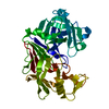
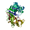
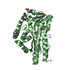
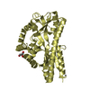
[English] 日本語
Yorodumi
- PDB-2cex: Structure of a sialic acid binding protein (SiaP) in the presence... -

+
Open data


ID or keywords:

Loading...

-
Basic information

Entry
Database: PDB / ID: 2cex
TitleStructure of a sialic acid binding protein (SiaP) in the presence of the sialic acid acid analogue Neu5Ac2en
ComponentsPROTEIN HI0146
KeywordsTRANSPORT / ESR / PERIPLASMIC BINDING PROTEIN / TRIPARTITE ATP-INDEPENDENT PERIPLASMIC (TRAP)TRANSPORT / VIRULENCE FACTOR / PERIPLASMIC
Function / homology
Function and homology information


: / transmembrane transport / outer membrane-bounded periplasmic space
Similarity search - Function
TRAP transporter solute receptor, DctP family / Bacterial extracellular solute-binding protein, family 7 / TRAP transporter solute receptor DctP / TRAP transporter solute receptor DctP superfamily / Bacterial extracellular solute-binding protein, family 7 / D-Maltodextrin-Binding Protein; domain 2 / 3-Layer(aba) Sandwich / Alpha Beta
Similarity search - Domain/homology
2-DEOXY-2,3-DEHYDRO-N-ACETYL-NEURAMINIC ACID / Sialic acid-binding periplasmic protein SiaP
Similarity search - Component
Biological speciesHAEMOPHILUS INFLUENZAE (bacteria)
MethodX-RAY DIFFRACTION / SYNCHROTRON / MOLECULAR REPLACEMENT / Resolution: 2.2 Å
AuthorsMuller, A. / Severi, E. / Mulligan, C. / Watts, A.G. / Kelly, D.J. / Wilson, K.S. / Wilkinson, A.J. / Thomas, G.H.
CitationJournal: J.Biol.Chem. / Year: 2006
Title: Conservation of Structure and Mechanism in Primary and Secondary Transporters Exemplified by Siap, a Sialic Acid Binding Virulence Factor from Haemophilus Influenzae
Authors: Muller, A. / Severi, E. / Mulligan, C. / Watts, A.G. / Kelly, D.J. / Wilson, K.S. / Wilkinson, A.J. / Thomas, G.H.
History
DepositionFeb 10, 2006Deposition site: PDBE / Processing site: PDBE
Revision 1.0May 15, 2006Provider: repository / Type: Initial release
Revision 1.1Jul 13, 2011Group: Advisory / Refinement description / Version format compliance
Revision 1.2Jul 29, 2020Group: Data collection / Derived calculations / Other
Category: chem_comp / pdbx_database_status ...chem_comp / pdbx_database_status / pdbx_struct_conn_angle / struct_conn / struct_site / struct_site_gen
Item: _chem_comp.type / _pdbx_database_status.status_code_sf ..._chem_comp.type / _pdbx_database_status.status_code_sf / _pdbx_struct_conn_angle.ptnr1_auth_asym_id / _pdbx_struct_conn_angle.ptnr1_auth_comp_id / _pdbx_struct_conn_angle.ptnr1_auth_seq_id / _pdbx_struct_conn_angle.ptnr1_label_asym_id / _pdbx_struct_conn_angle.ptnr1_label_atom_id / _pdbx_struct_conn_angle.ptnr1_label_comp_id / _pdbx_struct_conn_angle.ptnr1_label_seq_id / _pdbx_struct_conn_angle.ptnr2_auth_seq_id / _pdbx_struct_conn_angle.ptnr2_label_asym_id / _pdbx_struct_conn_angle.ptnr3_auth_asym_id / _pdbx_struct_conn_angle.ptnr3_auth_comp_id / _pdbx_struct_conn_angle.ptnr3_auth_seq_id / _pdbx_struct_conn_angle.ptnr3_label_asym_id / _pdbx_struct_conn_angle.ptnr3_label_atom_id / _pdbx_struct_conn_angle.ptnr3_label_comp_id / _pdbx_struct_conn_angle.ptnr3_label_seq_id / _pdbx_struct_conn_angle.value / _struct_conn.pdbx_dist_value / _struct_conn.ptnr1_auth_asym_id / _struct_conn.ptnr1_auth_comp_id / _struct_conn.ptnr1_auth_seq_id / _struct_conn.ptnr1_label_asym_id / _struct_conn.ptnr1_label_atom_id / _struct_conn.ptnr1_label_comp_id / _struct_conn.ptnr1_label_seq_id / _struct_conn.ptnr1_symmetry / _struct_conn.ptnr2_auth_asym_id / _struct_conn.ptnr2_auth_comp_id / _struct_conn.ptnr2_auth_seq_id / _struct_conn.ptnr2_label_asym_id / _struct_conn.ptnr2_label_atom_id / _struct_conn.ptnr2_label_comp_id / _struct_conn.ptnr2_label_seq_id / _struct_conn.ptnr2_symmetry
Description: Carbohydrate remediation / Provider: repository / Type: Remediation
Revision 1.3May 8, 2024Group: Data collection / Database references / Structure summary
Category: chem_comp / chem_comp_atom ...chem_comp / chem_comp_atom / chem_comp_bond / database_2
Item: _chem_comp.pdbx_synonyms / _database_2.pdbx_DOI / _database_2.pdbx_database_accession
Remark 700 SHEET THE SHEET STRUCTURE OF THIS MOLECULE IS BIFURCATED. IN ORDER TO REPRESENT THIS FEATURE IN ... SHEET THE SHEET STRUCTURE OF THIS MOLECULE IS BIFURCATED. IN ORDER TO REPRESENT THIS FEATURE IN THE SHEET RECORDS BELOW, TWO SHEETS ARE DEFINED.

-
Structure visualization

Structure viewerMolecule:
MolmilJmol/JSmol

Downloads & links

-
Assembly

Deposited unit
A: PROTEIN HI0146
B: PROTEIN HI0146
C: PROTEIN HI0146
D: PROTEIN HI0146
hetero molecules


Theoretical massNumber of molelcules
Total (without water)137,77514
Polymers136,8154
Non-polymers96010
Water8,197455
1
A: PROTEIN HI0146
hetero molecules


Theoretical massNumber of molelcules
Total (without water)34,2692
Polymers34,2041
Non-polymers651
Water181
TypeNameSymmetry operationNumber
identity operation1_555x,y,z1
MethodPISA
2
B: PROTEIN HI0146
hetero molecules


Theoretical massNumber of molelcules
Total (without water)34,8106
Polymers34,2041
Non-polymers6065
Water181
TypeNameSymmetry operationNumber
identity operation1_555x,y,z1
MethodPISA
3
C: PROTEIN HI0146
hetero molecules


Theoretical massNumber of molelcules
Total (without water)34,3353
Polymers34,2041
Non-polymers1312
Water181
TypeNameSymmetry operationNumber
identity operation1_555x,y,z1
MethodPISA
4
D: PROTEIN HI0146
hetero molecules


Theoretical massNumber of molelcules
Total (without water)34,3613
Polymers34,2041
Non-polymers1582
Water181
TypeNameSymmetry operationNumber
identity operation1_555x,y,z1
MethodPISA
Unit cell
Length a, b, c (Å)131.448, 88.701, 115.912
Angle α, β, γ (deg.)90.00, 105.33, 90.00
Int Tables number5
Space group name H-MC121

-
Components

#1: Protein
PROTEIN HI0146 / SIAP


Mass: 34203.746 Da / Num. of mol.: 4
Source method: isolated from a genetically manipulated source
Source: (gene. exp.) HAEMOPHILUS INFLUENZAE (bacteria) / Plasmid: PET21B / Production host: ESCHERICHIA COLI (E. coli) / Strain (production host): BL21(DE3) / References: UniProt: P44542
#2: Chemical
ChemComp-ZN / ZINC ION


Mass: 65.409 Da / Num. of mol.: 6 / Source method: obtained synthetically / Formula: Zn
#3: Sugar ChemComp-DAN / 2-DEOXY-2,3-DEHYDRO-N-ACETYL-NEURAMINIC ACID / Neu5Ac2en


Type: D-saccharide / Mass: 291.255 Da / Num. of mol.: 1 / Source method: obtained synthetically / Formula: C11H17NO8
#4: Chemical ChemComp-GOL / GLYCEROL / GLYCERIN / PROPANE-1,2,3-TRIOL / Glycerol


Mass: 92.094 Da / Num. of mol.: 3 / Source method: obtained synthetically / Formula: C3H8O3
#5: Water ChemComp-HOH / water / Water


Mass: 18.015 Da / Num. of mol.: 455 / Source method: isolated from a natural source / Formula: H2O

-
Experimental details

-
Experiment

ExperimentMethod: X-RAY DIFFRACTION / Number of used crystals: 1

-
Sample preparation

CrystalDensity Matthews: 2.32 Å3/Da / Density % sol: 46.5 %
Crystal growpH: 8.5
Details: 5 MM NEU5AC2EN, 5 MM ZINC ACETATE,0.1 M TRIS-HCL PH 8.5, 0.2 M MAGNESIUM CHLORIDE, 25 % PEG 3350

-
Data collection

DiffractionMean temperature: 100 K
Diffraction sourceSource: SYNCHROTRON / Site: ESRF / Beamline: BM14 / Wavelength: 0.97624
DetectorType: MARRESEARCH / Detector: CCD / Date: Jun 26, 2005
RadiationProtocol: SINGLE WAVELENGTH / Monochromatic (M) / Laue (L): M / Scattering type: x-ray
Radiation wavelengthWavelength: 0.97624 Å / Relative weight: 1
ReflectionResolution: 2.2→50 Å / Num. obs: 49350 / % possible obs: 75.7 % / Observed criterion σ(I): 3 / Redundancy: 3.2 % / Rmerge(I) obs: 0.1 / Net I/σ(I): 10.7
Reflection shellResolution: 2.2→2.28 Å / Redundancy: 1.3 % / Rmerge(I) obs: 0.56 / Mean I/σ(I) obs: 1.1 / % possible all: 12.7

-
Processing

Software
NameVersionClassification
REFMAC5.2.0005refinement
DENZOdata reduction
SCALEPACKdata scaling
MOLREPphasing
RefinementMethod to determine structure: MOLECULAR REPLACEMENT / Resolution: 2.2→111.8 Å / Cor.coef. Fo:Fc: 0.939 / Cor.coef. Fo:Fc free: 0.877 / SU B: 16.616 / SU ML: 0.239 / TLS residual ADP flag: LIKELY RESIDUAL / Cross valid method: THROUGHOUT / ESU R: 0.588 / ESU R Free: 0.319 / Stereochemistry target values: MAXIMUM LIKELIHOOD / Details: HYDROGENS HAVE BEEN ADDED IN THE RIDING POSITIONS
RfactorNum. reflection% reflectionSelection details
Rfree0.28 2500 5.1 %RANDOM
Rwork0.197 ---
obs0.201 46872 75.6 %-
Solvent computationIon probe radii: 0.8 Å / Shrinkage radii: 0.8 Å / VDW probe radii: 1.2 Å / Solvent model: MASK
Displacement parametersBiso mean: 30.03 Å2
Baniso -1Baniso -2Baniso -3
1-0.12 Å20 Å20.1 Å2
2--0.32 Å20 Å2
3----0.39 Å2
Refinement stepCycle: LAST / Resolution: 2.2→111.8 Å
ProteinNucleic acidLigandSolventTotal
Num. atoms9515 0 44 455 10014
Refine LS restraints
Refine-IDTypeDev idealDev ideal targetNumber
X-RAY DIFFRACTIONr_bond_refined_d0.0180.0229745
X-RAY DIFFRACTIONr_bond_other_d0.0010.028677
X-RAY DIFFRACTIONr_angle_refined_deg1.7271.97113156
X-RAY DIFFRACTIONr_angle_other_deg0.985320357
X-RAY DIFFRACTIONr_dihedral_angle_1_deg7.3751211
X-RAY DIFFRACTIONr_dihedral_angle_2_deg39.81425.928447
X-RAY DIFFRACTIONr_dihedral_angle_3_deg18.282151727
X-RAY DIFFRACTIONr_dihedral_angle_4_deg11.8051519
X-RAY DIFFRACTIONr_chiral_restr0.110.21456
X-RAY DIFFRACTIONr_gen_planes_refined0.0060.0210829
X-RAY DIFFRACTIONr_gen_planes_other0.0010.021865
X-RAY DIFFRACTIONr_nbd_refined0.1980.22330
X-RAY DIFFRACTIONr_nbd_other0.1820.29009
X-RAY DIFFRACTIONr_nbtor_refined0.1770.24651
X-RAY DIFFRACTIONr_nbtor_other0.0930.25407
X-RAY DIFFRACTIONr_xyhbond_nbd_refined0.1940.2524
X-RAY DIFFRACTIONr_xyhbond_nbd_other0.0150.21
X-RAY DIFFRACTIONr_metal_ion_refined0.1640.24
X-RAY DIFFRACTIONr_metal_ion_other
X-RAY DIFFRACTIONr_symmetry_vdw_refined0.1530.225
X-RAY DIFFRACTIONr_symmetry_vdw_other0.2220.274
X-RAY DIFFRACTIONr_symmetry_hbond_refined0.1680.218
X-RAY DIFFRACTIONr_symmetry_hbond_other
X-RAY DIFFRACTIONr_symmetry_metal_ion_refined
X-RAY DIFFRACTIONr_symmetry_metal_ion_other
X-RAY DIFFRACTIONr_mcbond_it0.6291.56307
X-RAY DIFFRACTIONr_mcbond_other0.1431.52452
X-RAY DIFFRACTIONr_mcangle_it0.99729703
X-RAY DIFFRACTIONr_mcangle_other
X-RAY DIFFRACTIONr_scbond_it1.67834035
X-RAY DIFFRACTIONr_scbond_other
X-RAY DIFFRACTIONr_scangle_it2.4924.53453
X-RAY DIFFRACTIONr_scangle_other
X-RAY DIFFRACTIONr_long_range_B_refined
X-RAY DIFFRACTIONr_long_range_B_other
X-RAY DIFFRACTIONr_rigid_bond_restr
X-RAY DIFFRACTIONr_sphericity_free
X-RAY DIFFRACTIONr_sphericity_bonded
LS refinement shellResolution: 2.2→2.26 Å / Total num. of bins used: 20
RfactorNum. reflection% reflection
Rfree0.538 32 -
Rwork0.305 519 -
obs--11.48 %
Refinement TLS params.

Method: refined / Refine-ID: X-RAY DIFFRACTION

IDL112)L122)L132)L222)L232)L332)S11 (Å °)S12 (Å °)S13 (Å °)S21 (Å °)S22 (Å °)S23 (Å °)S31 (Å °)S32 (Å °)S33 (Å °)T112)T122)T132)T222)T232)T332)Origin x (Å)Origin y (Å)Origin z (Å)
12.470.73420.31192.34821.63973.1825-0.14340.5214-0.3669-0.36480.2476-0.1980.02240.2084-0.1043-0.0156-0.03760.0120.0907-0.0824-0.054221.413122.80585.1413
21.33390.5987-0.03731.2433-0.46112.052-0.1345-0.1668-0.0784-0.08310.0471-0.20920.08620.27160.0874-0.1334-0.01470.01840.0595-0.0754-0.059829.131334.014729.4307
31.9184-0.0403-0.08012.12950.17771.4042-0.04960.21230.2263-0.19210.04950.0951-0.1736-0.13490.0001-0.1417-0.02520.0083-0.03370.0303-0.127355.7239.27217.922
42.38320.33490.64271.62560.51181.1658-0.0048-0.13250.05940.16370.017-0.03460.0461-0.0429-0.0121-0.1447-0.01730.0227-0.0417-0.03-0.108263.7782-4.035634.3025
53.15871.3037-0.58020.7805-0.11612.3253-0.3140.8929-0.374-0.25130.2326-0.07580.2737-0.43710.0814-0.0954-0.13010.02890.2432-0.0763-0.003724.8345-15.526924.6442
62.18740.6629-0.81491.0202-1.07462.53310.0142-0.0111-0.0928-0.0366-0.0338-0.12910.01590.20360.0197-0.12940.0069-0.0084-0.0141-0.0225-0.083430.9496-5.264248.082
70.9770.06940.0421.6865-0.77561.0796-0.0625-0.00550.0878-0.16810.02880.15820.0234-0.09160.0337-0.0855-0.02370.001-0.1015-0.0058-0.066-4.68943.723830.6403
81.58040.2440.68710.48380.69582.6641-0.0162-0.34160.070.1093-0.08170.18350.0247-0.31060.0979-0.0949-0.00620.0329-0.01480.0569-0.0809-0.8558-7.089254.8517
Refinement TLS group
IDRefine-IDRefine TLS-IDAuth asym-IDAuth seq-ID
1X-RAY DIFFRACTION1A2 - 125
2X-RAY DIFFRACTION1A213 - 254
3X-RAY DIFFRACTION2A126 - 210
4X-RAY DIFFRACTION2A256 - 288
5X-RAY DIFFRACTION3B2 - 125
6X-RAY DIFFRACTION3B213 - 254
7X-RAY DIFFRACTION4B126 - 210
8X-RAY DIFFRACTION4B256 - 300
9X-RAY DIFFRACTION5C2 - 125
10X-RAY DIFFRACTION5C213 - 254
11X-RAY DIFFRACTION6C126 - 210
12X-RAY DIFFRACTION6C256 - 300
13X-RAY DIFFRACTION7D2 - 125
14X-RAY DIFFRACTION7D213 - 254
15X-RAY DIFFRACTION8D126 - 210
16X-RAY DIFFRACTION8D256 - 300

+
About Yorodumi

-
News

-
Feb 9, 2022. New format data for meta-information of EMDB entries

New format data for meta-information of EMDB entries

  • Version 3 of the EMDB header file is now the official format.
  • The previous official version 1.9 will be removed from the archive.

Related info.:EMDB header

External links:wwPDB to switch to version 3 of the EMDB data model

-
Aug 12, 2020. Covid-19 info

Covid-19 info

URL: https://pdbj.org/emnavi/covid19.php

New page: Covid-19 featured information page in EM Navigator.

Related info.:Covid-19 info / Mar 5, 2020. Novel coronavirus structure data

+
Mar 5, 2020. Novel coronavirus structure data

Novel coronavirus structure data

Related info.:Yorodumi Speices / Aug 12, 2020. Covid-19 info

External links:COVID-19 featured content - PDBj / Molecule of the Month (242):Coronavirus Proteases

+
Jan 31, 2019. EMDB accession codes are about to change! (news from PDBe EMDB page)

EMDB accession codes are about to change! (news from PDBe EMDB page)

  • The allocation of 4 digits for EMDB accession codes will soon come to an end. Whilst these codes will remain in use, new EMDB accession codes will include an additional digit and will expand incrementally as the available range of codes is exhausted. The current 4-digit format prefixed with “EMD-” (i.e. EMD-XXXX) will advance to a 5-digit format (i.e. EMD-XXXXX), and so on. It is currently estimated that the 4-digit codes will be depleted around Spring 2019, at which point the 5-digit format will come into force.
  • The EM Navigator/Yorodumi systems omit the EMD- prefix.

Related info.:Q: What is EMD? / ID/Accession-code notation in Yorodumi/EM Navigator

External links:EMDB Accession Codes are Changing Soon! / Contact to PDBj

+
Jul 12, 2017. Major update of PDB

Major update of PDB

  • wwPDB released updated PDB data conforming to the new PDBx/mmCIF dictionary.
  • This is a major update changing the version number from 4 to 5, and with Remediation, in which all the entries are updated.
  • In this update, many items about electron microscopy experimental information are reorganized (e.g. em_software).
  • Now, EM Navigator and Yorodumi are based on the updated data.

External links:wwPDB Remediation / Enriched Model Files Conforming to OneDep Data Standards Now Available in the PDB FTP Archive

-
Yorodumi

Thousand views of thousand structures

  • Yorodumi is a browser for structure data from EMDB, PDB, SASBDB, etc.
  • This page is also the successor to EM Navigator detail page, and also detail information page/front-end page for Omokage search.
  • The word "yorodu" (or yorozu) is an old Japanese word meaning "ten thousand". "mi" (miru) is to see.

Related info.:EMDB / PDB / SASBDB / Comparison of 3 databanks / Yorodumi Search / Aug 31, 2016. New EM Navigator & Yorodumi / Yorodumi Papers / Jmol/JSmol / Function and homology information / Changes in new EM Navigator and Yorodumi

Read more