 Movie
Movie Controller


+ Open data

- Basic information
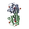
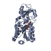
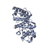
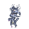
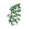
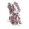
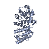
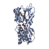
| Entry | Database: PDB / ID: 2xws | ||||||
|---|---|---|---|---|---|---|---|
| Title | ANAEROBIC COBALT CHELATASE (CbiX) FROM ARCHAEOGLOBUS FULGIDUS | ||||||
 Components Components | SIROHYDROCHLORIN COBALTOCHELATASE | ||||||
 Keywords Keywords | LYASE / BETA-ALPHA-BETA / COBALAMIN BIOSYNTHESIS / METAL-BINDING / PARALLEL BETA SHEET | ||||||
| Function / homology |  Function and homology informationsirohydrochlorin cobaltochelatase / anaerobic cobalamin biosynthetic process / sirohydrochlorin cobaltochelatase activity / tetrapyrrole binding / cobalt ion binding Function and homology informationsirohydrochlorin cobaltochelatase / anaerobic cobalamin biosynthetic process / sirohydrochlorin cobaltochelatase activity / tetrapyrrole binding / cobalt ion bindingSimilarity search - Function | ||||||
| Biological species |   ARCHAEOGLOBUS FULGIDUS (archaea) ARCHAEOGLOBUS FULGIDUS (archaea) | ||||||
| Method | X-RAY DIFFRACTION / SYNCHROTRON / MOLECULAR REPLACEMENT / Resolution: 1.6 Å | ||||||
 Authors Authors | Romao, C.V. / Ladakis, D. / Lobo, S.A.L. / Carrondo, M.A. / Brindley, A.A. / Deery, E. / Matias, P.M. / Pickersgill, R.W. / Saraiva, L.M. / Warren, M.J. | ||||||
 Citation Citation | Journal: Proc.Natl.Acad.Sci.USA / Year: 2011 Title: Evolution in a Family of Chelatases Facilitated by the Introduction of Active Site Asymmetry and Protein Oligomerization. Authors: Romao, C.V. / Ladakis, D. / Lobo, S.A. / Carrondo, M.A. / Brindley, A.A. / Deery, E. / Matias, P.M. / Pickersgill, R.W. / Saraiva, L.M. / Warren, M.J. | ||||||
| History |
|
- Structure visualization
| Structure viewer | Molecule: MolmilJmol/JSmol |
|---|
- Downloads & links
-Download
| PDBx/mmCIF format | 2xws.cif.gz | 40.1 KB | Display | PDBx/mmCIF format |
|---|---|---|---|---|
| PDB format | pdb2xws.ent.gz | 27.4 KB | Display | PDB format |
| PDBx/mmJSON format | 2xws.json.gz | Tree view | PDBx/mmJSON format | |
| Others |  Other downloads Other downloads |
-Validation report
| Arichive directory | https://data.pdbj.org/pub/pdb/validation_reports/xw/2xwsftp://data.pdbj.org/pub/pdb/validation_reports/xw/2xws | HTTPS FTP |
|---|
-Related structure data
| Related structure data |  2xvxC  2xvzC  2xwpC  2xwqC  2dj5S S: Starting model for refinement C: citing same article ( |
|---|---|
| Similar structure data |


-Links
 PDBj
PDBj- Assembly
| Deposited unit | 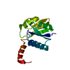
| ||||||||
|---|---|---|---|---|---|---|---|---|---|
| 1 | 
| ||||||||
| Unit cell |
|
-Components
| #1: Protein | Mass: 15190.501 Da / Num. of mol.: 1 Source method: isolated from a genetically manipulated source Source: (gene. exp.) ARCHAEOGLOBUS FULGIDUS (archaea) / Plasmid: PET3A / Production host:  ESCHERICHIA COLI (E. coli) / Strain (production host): BL21 STAR DE3 / Variant (production host): PLYSS ESCHERICHIA COLI (E. coli) / Strain (production host): BL21 STAR DE3 / Variant (production host): PLYSSReferences: UniProt: O29537, sirohydrochlorin cobaltochelatase |
|---|---|
| #2: Water | ChemComp-HOH / Water Mass: 18.015 Da / Num. of mol.: 88 / Source method: isolated from a natural source / Formula: H2O Mass: 18.015 Da / Num. of mol.: 88 / Source method: isolated from a natural source / Formula: H2O |
-Experimental details
-Experiment
| Experiment | Method: X-RAY DIFFRACTION / Number of used crystals: 1 |
|---|
- Sample preparation
| Crystal | Density Matthews: 2.23 Å3/Da / Density % sol: 45 % / Description: NONE |
|---|---|
| Crystal grow | pH: 6.8 / Details: pH 6.8 |
-Data collection
| Diffraction | Mean temperature: 100 K |
|---|---|
| Diffraction source | Source: SYNCHROTRON / Site: Diamond  / Beamline: I04 / Wavelength: 0.9835 / Beamline: I04 / Wavelength: 0.9835 |
| Detector | Type: ADSC CCD / Detector: CCD / Details: MIRRORS |
| Radiation | Monochromator: SILICON MONOCHROMATOR / Protocol: SINGLE WAVELENGTH / Monochromatic (M) / Laue (L): M / Scattering type: x-ray |
| Radiation wavelength | Wavelength: 0.9835 Å / Relative weight: 1 |
| Reflection | Resolution: 1.6→29.5 Å / Num. obs: 118464 / % possible obs: 99.4 % / Observed criterion σ(I): 0 / Redundancy: 6.4 % / Biso Wilson estimate: 13.6 Å2 / Rmerge(I) obs: 0.05 / Net I/σ(I): 23.6 |
| Reflection shell | Resolution: 1.6→1.69 Å / Redundancy: 6.7 % / Rmerge(I) obs: 0.13 / Mean I/σ(I) obs: 13.3 / % possible all: 100 |
- Processing
| Software |
| ||||||||||||||||||||||||||||||||||||||||||||||||||||||||||||||||||||||||||||||||||||||||||||||||||||||||||||||||||||||||||||||||||||||||||||||||||||||||||||||||||||||||||||||||||||||
|---|---|---|---|---|---|---|---|---|---|---|---|---|---|---|---|---|---|---|---|---|---|---|---|---|---|---|---|---|---|---|---|---|---|---|---|---|---|---|---|---|---|---|---|---|---|---|---|---|---|---|---|---|---|---|---|---|---|---|---|---|---|---|---|---|---|---|---|---|---|---|---|---|---|---|---|---|---|---|---|---|---|---|---|---|---|---|---|---|---|---|---|---|---|---|---|---|---|---|---|---|---|---|---|---|---|---|---|---|---|---|---|---|---|---|---|---|---|---|---|---|---|---|---|---|---|---|---|---|---|---|---|---|---|---|---|---|---|---|---|---|---|---|---|---|---|---|---|---|---|---|---|---|---|---|---|---|---|---|---|---|---|---|---|---|---|---|---|---|---|---|---|---|---|---|---|---|---|---|---|---|---|---|---|
| Refinement | Method to determine structure: MOLECULAR REPLACEMENT Starting model: PDB ENTRY 2DJ5 Resolution: 1.6→33.99 Å / Cor.coef. Fo:Fc: 0.954 / Cor.coef. Fo:Fc free: 0.943 / SU B: 1.475 / SU ML: 0.053 / Cross valid method: THROUGHOUT / ESU R: 0.088 / ESU R Free: 0.088 / Stereochemistry target values: MAXIMUM LIKELIHOOD Details: HYDROGENS HAVE BEEN ADDED IN THE RIDING POSITIONS. U VALUES REFINED INDIVIDUALLY.
| ||||||||||||||||||||||||||||||||||||||||||||||||||||||||||||||||||||||||||||||||||||||||||||||||||||||||||||||||||||||||||||||||||||||||||||||||||||||||||||||||||||||||||||||||||||||
| Solvent computation | Ion probe radii: 0.8 Å / Shrinkage radii: 0.8 Å / VDW probe radii: 1.4 Å / Solvent model: MASK | ||||||||||||||||||||||||||||||||||||||||||||||||||||||||||||||||||||||||||||||||||||||||||||||||||||||||||||||||||||||||||||||||||||||||||||||||||||||||||||||||||||||||||||||||||||||
| Displacement parameters | Biso mean: 16.203 Å2
| ||||||||||||||||||||||||||||||||||||||||||||||||||||||||||||||||||||||||||||||||||||||||||||||||||||||||||||||||||||||||||||||||||||||||||||||||||||||||||||||||||||||||||||||||||||||
| Refinement step | Cycle: LAST / Resolution: 1.6→33.99 Å
| ||||||||||||||||||||||||||||||||||||||||||||||||||||||||||||||||||||||||||||||||||||||||||||||||||||||||||||||||||||||||||||||||||||||||||||||||||||||||||||||||||||||||||||||||||||||
| Refine LS restraints |
|