 Movie
Movie Controller

[English] 日本語
 Yorodumi
- PDB-2xta: Crystal structure of the SucA domain of Mycobacterium smegmatis a... -
+ Open data

- Basic information
| Entry | Database: PDB / ID: 2xta | ||||||
|---|---|---|---|---|---|---|---|
| Title | Crystal structure of the SucA domain of Mycobacterium smegmatis alpha- ketoglutarate decarboxylase in complex with acetyl-CoA (triclinic form) | ||||||
 Components Components | 2-OXOGLUTARATE DECARBOXYLASE | ||||||
 Keywords Keywords | LYASE / KDH / KGD | ||||||
| Function / homology |  Function and homology information2-hydroxy-3-oxoadipate synthase / 2-oxoglutarate decarboxylase / 2-oxoglutarate decarboxylase activity / 2-hydroxy-3-oxoadipate synthase activity / oxoglutarate dehydrogenase (succinyl-transferring) / oxoglutarate dehydrogenase (succinyl-transferring) activity / dihydrolipoyllysine-residue succinyltransferase / dihydrolipoyllysine-residue succinyltransferase activity / oxoglutarate dehydrogenase complex / thiamine pyrophosphate binding ...2-hydroxy-3-oxoadipate synthase / 2-oxoglutarate decarboxylase / 2-oxoglutarate decarboxylase activity / 2-hydroxy-3-oxoadipate synthase activity / oxoglutarate dehydrogenase (succinyl-transferring) / oxoglutarate dehydrogenase (succinyl-transferring) activity / dihydrolipoyllysine-residue succinyltransferase / dihydrolipoyllysine-residue succinyltransferase activity / oxoglutarate dehydrogenase complex / thiamine pyrophosphate binding / tricarboxylic acid cycle / magnesium ion binding / cytosol Function and homology information2-hydroxy-3-oxoadipate synthase / 2-oxoglutarate decarboxylase / 2-oxoglutarate decarboxylase activity / 2-hydroxy-3-oxoadipate synthase activity / oxoglutarate dehydrogenase (succinyl-transferring) / oxoglutarate dehydrogenase (succinyl-transferring) activity / dihydrolipoyllysine-residue succinyltransferase / dihydrolipoyllysine-residue succinyltransferase activity / oxoglutarate dehydrogenase complex / thiamine pyrophosphate binding ...2-hydroxy-3-oxoadipate synthase / 2-oxoglutarate decarboxylase / 2-oxoglutarate decarboxylase activity / 2-hydroxy-3-oxoadipate synthase activity / oxoglutarate dehydrogenase (succinyl-transferring) / oxoglutarate dehydrogenase (succinyl-transferring) activity / dihydrolipoyllysine-residue succinyltransferase / dihydrolipoyllysine-residue succinyltransferase activity / oxoglutarate dehydrogenase complex / thiamine pyrophosphate binding / tricarboxylic acid cycle / magnesium ion binding / cytosolSimilarity search - Function | ||||||
| Biological species |  MYCOBACTERIUM SMEGMATIS (bacteria) MYCOBACTERIUM SMEGMATIS (bacteria) | ||||||
| Method | X-RAY DIFFRACTION / SYNCHROTRON / MOLECULAR REPLACEMENT / Resolution: 2.2 Å | ||||||
 Authors Authors | Wagner, T. / Bellinzoni, M. / Wehenkel, A.M. / O'Hare, H.M. / Alzari, P.M. | ||||||
 Citation Citation | Journal: Chem.Biol. / Year: 2011 Title: Functional Plasticity and Allosteric Regulation of Alpha-Ketoglutarate Decarboxylase in Central Mycobacterial Metabolism. Authors: Wagner, T. / Bellinzoni, M. / Wehenkel, A.M. / O'Hare, H.M. / Alzari, P.M. | ||||||
| History |
|
- Structure visualization
| Structure viewer | Molecule: MolmilJmol/JSmol |
|---|
- Downloads & links
-Download
| PDBx/mmCIF format | 2xta.cif.gz | 1.3 MB | Display | PDBx/mmCIF format |
|---|---|---|---|---|
| PDB format | pdb2xta.ent.gz | 1 MB | Display | PDB format |
| PDBx/mmJSON format | 2xta.json.gz | Tree view | PDBx/mmJSON format | |
| Others |  Other downloads Other downloads |
-Validation report
| Arichive directory | https://data.pdbj.org/pub/pdb/validation_reports/xt/2xtaftp://data.pdbj.org/pub/pdb/validation_reports/xt/2xta | HTTPS FTP |
|---|
-Related structure data
| Related structure data | 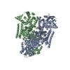 2xt6C  2y0pC 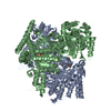 2yicC  2yidC  2xt9 2xt5 2xt7 2xt8 C: citing same article ( S: Starting model for refinement |
|---|---|
| Similar structure data |


-Links
 PDBj
PDBj


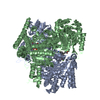
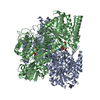
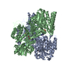
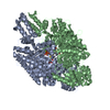
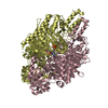
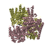
- Assembly
| Deposited unit | 
| ||||||||||||||||
|---|---|---|---|---|---|---|---|---|---|---|---|---|---|---|---|---|---|
| 1 | 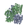
| ||||||||||||||||
| 2 | 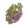
| ||||||||||||||||
| Unit cell |
| ||||||||||||||||
| Noncrystallographic symmetry (NCS) | NCS oper:
|
-Components
-Protein , 1 types, 4 molecules ABCD
| #1: Protein | / ALPHA-KETOGLUTARATE DECARBOXYLASE / 2-OXOGLUTARATE CARBOXY-LYASE Mass: 97166.648 Da / Num. of mol.: 4 / Fragment: RESIDUES 361-1227 Source method: isolated from a genetically manipulated source Source: (gene. exp.) MYCOBACTERIUM SMEGMATIS (bacteria) / Strain: MC2 155 / Plasmid: PET-28A / Production host: ESCHERICHIA COLI (E. coli) / Strain (production host): BL21(DE3) PLYSS / References: UniProt: A0R2B1, 2-oxoglutarate decarboxylase |
|---|
-Non-polymers , 5 types, 757 molecules 


| #2: Chemical | ChemComp-TPP / Thiamine pyrophosphate Mass: 425.314 Da / Num. of mol.: 4 / Source method: obtained synthetically / Formula: C12H19N4O7P2S Mass: 425.314 Da / Num. of mol.: 4 / Source method: obtained synthetically / Formula: C12H19N4O7P2S#3: Chemical | ChemComp-MG /  Mass: 24.305 Da / Num. of mol.: 4 / Source method: obtained synthetically / Formula: Mg Mass: 24.305 Da / Num. of mol.: 4 / Source method: obtained synthetically / Formula: Mg#4: Chemical | ChemComp-CA /  Mass: 40.078 Da / Num. of mol.: 4 / Source method: obtained synthetically / Formula: Ca Mass: 40.078 Da / Num. of mol.: 4 / Source method: obtained synthetically / Formula: Ca#5: Chemical | Acetyl-CoA Mass: 809.571 Da / Num. of mol.: 3 / Source method: obtained synthetically / Formula: C23H38N7O17P3S Mass: 809.571 Da / Num. of mol.: 3 / Source method: obtained synthetically / Formula: C23H38N7O17P3S#6: Water | ChemComp-HOH / | WaterMass: 18.015 Da / Num. of mol.: 742 / Source method: isolated from a natural source / Formula: H2O |
|---|
-Experimental details
-Experiment
| Experiment | Method: X-RAY DIFFRACTION / Number of used crystals: 1 |
|---|
- Sample preparation
| Crystal | Density Matthews: 2.79 Å3/Da / Density % sol: 55.9 % / Description: NONE |
|---|---|
| Crystal grow | pH: 7.6 / Details: 54% MPD, 25 MM SODIUM ACETATE, pH 7.6 |
-Data collection
| Diffraction | Mean temperature: 100 K |
|---|---|
| Diffraction source | Source: SYNCHROTRON / Site: SLS  / Beamline: X06DA / Wavelength: 0.9799 / Beamline: X06DA / Wavelength: 0.9799 |
| Detector | Type: MARMOSAIC 225 mm CCD / Detector: CCD / Date: Nov 21, 2009 / Details: TOROIDAL MIRROR |
| Radiation | Monochromator: DUAL CHANNEL CUT CRYSTALS / Protocol: SINGLE WAVELENGTH / Monochromatic (M) / Laue (L): M / Scattering type: x-ray |
| Radiation wavelength | Wavelength: 0.9799 Å / Relative weight: 1 |
| Reflection | Resolution: 2.2→78.1 Å / Num. obs: 187791 / % possible obs: 92.6 % / Observed criterion σ(I): 2.2 / Redundancy: 2 % / Biso Wilson estimate: 36.62 Å2 / Rmerge(I) obs: 0.07 / Net I/σ(I): 8.7 |
| Reflection shell | Resolution: 2.2→2.31 Å / Redundancy: 2 % / Rmerge(I) obs: 0.4 / Mean I/σ(I) obs: 2.2 / % possible all: 88.5 |
- Processing
| Software |
| |||||||||||||||||||||||||||||||||||||||||||||||||||||||||||||||||||||||||||||||||||||||||||||||||||||||||||||||||||||||||||||
|---|---|---|---|---|---|---|---|---|---|---|---|---|---|---|---|---|---|---|---|---|---|---|---|---|---|---|---|---|---|---|---|---|---|---|---|---|---|---|---|---|---|---|---|---|---|---|---|---|---|---|---|---|---|---|---|---|---|---|---|---|---|---|---|---|---|---|---|---|---|---|---|---|---|---|---|---|---|---|---|---|---|---|---|---|---|---|---|---|---|---|---|---|---|---|---|---|---|---|---|---|---|---|---|---|---|---|---|---|---|---|---|---|---|---|---|---|---|---|---|---|---|---|---|---|---|---|
| Refinement | Method to determine structure: MOLECULAR REPLACEMENT Starting model: PDB ENTRY 2XT9 2xt9 Resolution: 2.2→78.11 Å / Cor.coef. Fo:Fc: 0.939 / Cor.coef. Fo:Fc free: 0.9207 / SU R Cruickshank DPI: 0.236 / Cross valid method: THROUGHOUT / σ(F): 0 / SU R Blow DPI: 0.223 / SU Rfree Blow DPI: 0.173 / SU Rfree Cruickshank DPI: 0.179
| |||||||||||||||||||||||||||||||||||||||||||||||||||||||||||||||||||||||||||||||||||||||||||||||||||||||||||||||||||||||||||||
| Displacement parameters | Biso mean: 43.83 Å2
| |||||||||||||||||||||||||||||||||||||||||||||||||||||||||||||||||||||||||||||||||||||||||||||||||||||||||||||||||||||||||||||
| Refine analyze | Luzzati coordinate error obs: 0.299 Å | |||||||||||||||||||||||||||||||||||||||||||||||||||||||||||||||||||||||||||||||||||||||||||||||||||||||||||||||||||||||||||||
| Refinement step | Cycle: LAST / Resolution: 2.2→78.11 Å
| |||||||||||||||||||||||||||||||||||||||||||||||||||||||||||||||||||||||||||||||||||||||||||||||||||||||||||||||||||||||||||||
| Refine LS restraints |
| |||||||||||||||||||||||||||||||||||||||||||||||||||||||||||||||||||||||||||||||||||||||||||||||||||||||||||||||||||||||||||||
| LS refinement shell | Resolution: 2.2→2.26 Å / Total num. of bins used: 20
| |||||||||||||||||||||||||||||||||||||||||||||||||||||||||||||||||||||||||||||||||||||||||||||||||||||||||||||||||||||||||||||
| Refinement TLS params. | Method: refined / Refine-ID: X-RAY DIFFRACTION
| |||||||||||||||||||||||||||||||||||||||||||||||||||||||||||||||||||||||||||||||||||||||||||||||||||||||||||||||||||||||||||||
| Refinement TLS group |
|
