 Movie
Movie Controller


+ Open data

- Basic information
| Entry | Database: PDB / ID: 2xkl | ||||||
|---|---|---|---|---|---|---|---|
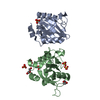
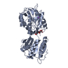
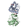
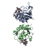
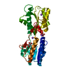
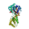
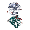
| Title | Crystal Structure of Mouse Apolipoprotein M | ||||||
 Components Components | APOLIPOPROTEIN M APOM APOM | ||||||
 Keywords Keywords | LIPID TRANSPORT | ||||||
| Function / homology |  Function and homology information Function and homology informationnegative regulation of plasma lipoprotein oxidation / Retinoid metabolism and transport / spherical high-density lipoprotein particle / discoidal high-density lipoprotein particle / lipid transporter activity / lipoprotein metabolic process / high-density lipoprotein particle remodeling / high-density lipoprotein particle clearance / low-density lipoprotein particle / reverse cholesterol transport ...negative regulation of plasma lipoprotein oxidation / Retinoid metabolism and transport / spherical high-density lipoprotein particle / discoidal high-density lipoprotein particle / lipid transporter activity / lipoprotein metabolic process / high-density lipoprotein particle remodeling / high-density lipoprotein particle clearance / low-density lipoprotein particle / reverse cholesterol transport / high-density lipoprotein particle assembly / very-low-density lipoprotein particle / high-density lipoprotein particle / cholesterol efflux / antioxidant activity / response to glucose / phospholipid binding / extracellular space / extracellular regionSimilarity search - Function | ||||||
| Biological species |  MUS MUSCULUS (house mouse) MUS MUSCULUS (house mouse) | ||||||
| Method | X-RAY DIFFRACTION / SYNCHROTRON / MOLECULAR REPLACEMENT / Resolution: 2.5 Å | ||||||
 Authors Authors | Sevvana, M. / Kassler, K. / Josefin, A. / Weiler, S. / Dahlback, B. / Sticht, H. / Muller, Y.A. | ||||||
 Citation Citation | Journal: J.Mol.Biol. / Year: 2010 Title: Mouse Apom Displays an Unprecedented Seven-Stranded Lipocalin Fold: Folding Decoy or Alternative Native Fold? Authors: Sevvana, M. / Kassler, K. / Ahnstrom, J. / Weiler, S. / Dahlback, B. / Sticht, H. / Muller, Y.A. | ||||||
| History |
|
- Structure visualization
| Structure viewer | Molecule: MolmilJmol/JSmol |
|---|
- Downloads & links
-Download
| PDBx/mmCIF format | 2xkl.cif.gz | 76.4 KB | Display | PDBx/mmCIF format |
|---|---|---|---|---|
| PDB format | pdb2xkl.ent.gz | 56.1 KB | Display | PDB format |
| PDBx/mmJSON format | 2xkl.json.gz | Tree view | PDBx/mmJSON format | |
| Others |  Other downloads Other downloads |
-Validation report
| Arichive directory | https://data.pdbj.org/pub/pdb/validation_reports/xk/2xklftp://data.pdbj.org/pub/pdb/validation_reports/xk/2xkl | HTTPS FTP |
|---|
-Related structure data
| Related structure data |  2wewS S: Starting model for refinement |
|---|---|
| Similar structure data |


-Links
 PDBj
PDBj

- Assembly
| Deposited unit | 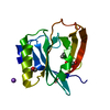
| ||||||||
|---|---|---|---|---|---|---|---|---|---|
| 1 | 
| ||||||||
| Unit cell |
|
-Components
-Protein , 1 types, 1 molecules A
| #1: Protein | APOM / APOM / APO-M Mass: 19101.688 Da / Num. of mol.: 1 / Fragment: RESIDUES 20-190 Source method: isolated from a genetically manipulated source Source: (gene. exp.) MUS MUSCULUS (house mouse) / Plasmid: PET30XA/LIC / Production host:  ESCHERICHIA COLI (E. coli) / Strain (production host): BL21 / References: UniProt: Q9Z1R3 ESCHERICHIA COLI (E. coli) / Strain (production host): BL21 / References: UniProt: Q9Z1R3 |
|---|
-Non-polymers , 5 types, 97 molecules 


| #2: Chemical | ChemComp-GOL / Glycerol Mass: 92.094 Da / Num. of mol.: 1 / Source method: obtained synthetically / Formula: C3H8O3 Mass: 92.094 Da / Num. of mol.: 1 / Source method: obtained synthetically / Formula: C3H8O3 |
|---|---|
| #3: Chemical | ChemComp-POL / Propan-1-ol Mass: 60.095 Da / Num. of mol.: 1 / Source method: obtained synthetically / Formula: C3H8O Mass: 60.095 Da / Num. of mol.: 1 / Source method: obtained synthetically / Formula: C3H8O |
| #4: Chemical | ChemComp-EDO / Ethylene glycol Mass: 62.068 Da / Num. of mol.: 1 / Source method: obtained synthetically / Formula: C2H6O2 Mass: 62.068 Da / Num. of mol.: 1 / Source method: obtained synthetically / Formula: C2H6O2 |
| #5: Chemical | ChemComp-NA /  Mass: 22.990 Da / Num. of mol.: 1 / Source method: obtained synthetically / Formula: Na Mass: 22.990 Da / Num. of mol.: 1 / Source method: obtained synthetically / Formula: Na |
| #6: Water | ChemComp-HOH / WaterMass: 18.015 Da / Num. of mol.: 93 / Source method: isolated from a natural source / Formula: H2O |
-Experimental details
-Experiment
| Experiment | Method: X-RAY DIFFRACTION / Number of used crystals: 1 |
|---|
- Sample preparation
| Crystal | Density Matthews: 2.28 Å3/Da / Density % sol: 46.02 % / Description: NONE |
|---|---|
| Crystal grow | pH: 5.6 Details: 0.1 M NA CITRATE PH 5.6, 20% V/V 2-PROPANOL, 20% W/V PEG 4000 |
-Data collection
| Diffraction | Mean temperature: 100 K |
|---|---|
| Diffraction source | Source: SYNCHROTRON / Site: BESSY  / Type: BESSY / Wavelength: 0.9184 / Type: BESSY / Wavelength: 0.9184 |
| Detector | Type: MARRESEARCH / Detector: CCD |
| Radiation | Protocol: SINGLE WAVELENGTH / Monochromatic (M) / Laue (L): M / Scattering type: x-ray |
| Radiation wavelength | Wavelength: 0.9184 Å / Relative weight: 1 |
| Reflection | Resolution: 2.5→20 Å / Num. obs: 6802 / % possible obs: 99.7 % / Observed criterion σ(I): 2 / Redundancy: 7.7 % / Biso Wilson estimate: 26.489 Å2 / Rmerge(I) obs: 0.18 / Net I/σ(I): 10.84 |
| Reflection shell | Resolution: 2.5→2.65 Å / Redundancy: 8.08 % / Rmerge(I) obs: 0.58 / Mean I/σ(I) obs: 3.56 / % possible all: 99.9 |
- Processing
| Software |
| ||||||||||||||||||||||||||||||||||||||||||||||||||||||||||||||||||||||||||||||||||||||||||||||||||||||||||||||||||||||||||||||||||||||||||||||||||||||||||||||||||||||||||||||||||||||
|---|---|---|---|---|---|---|---|---|---|---|---|---|---|---|---|---|---|---|---|---|---|---|---|---|---|---|---|---|---|---|---|---|---|---|---|---|---|---|---|---|---|---|---|---|---|---|---|---|---|---|---|---|---|---|---|---|---|---|---|---|---|---|---|---|---|---|---|---|---|---|---|---|---|---|---|---|---|---|---|---|---|---|---|---|---|---|---|---|---|---|---|---|---|---|---|---|---|---|---|---|---|---|---|---|---|---|---|---|---|---|---|---|---|---|---|---|---|---|---|---|---|---|---|---|---|---|---|---|---|---|---|---|---|---|---|---|---|---|---|---|---|---|---|---|---|---|---|---|---|---|---|---|---|---|---|---|---|---|---|---|---|---|---|---|---|---|---|---|---|---|---|---|---|---|---|---|---|---|---|---|---|---|---|
| Refinement | Method to determine structure: MOLECULAR REPLACEMENT Starting model: PDB ENTRY 2WEW Resolution: 2.5→19.94 Å / Cor.coef. Fo:Fc: 0.936 / Cor.coef. Fo:Fc free: 0.88 / SU B: 16.247 / SU ML: 0.195 / Cross valid method: THROUGHOUT / ESU R: 0.46 / ESU R Free: 0.279 / Stereochemistry target values: MAXIMUM LIKELIHOOD Details: HYDROGENS HAVE BEEN ADDED IN THE RIDING POSITIONS. ATOM RECORD CONTAINS SUM OF TLS AND RESIDUAL B FACTORS. ANISOU RECORD CONTAINS SUM OF TLS AND RESIDUAL U FACTORS.
| ||||||||||||||||||||||||||||||||||||||||||||||||||||||||||||||||||||||||||||||||||||||||||||||||||||||||||||||||||||||||||||||||||||||||||||||||||||||||||||||||||||||||||||||||||||||
| Solvent computation | Ion probe radii: 0.8 Å / Shrinkage radii: 0.8 Å / VDW probe radii: 1.2 Å / Solvent model: MASK | ||||||||||||||||||||||||||||||||||||||||||||||||||||||||||||||||||||||||||||||||||||||||||||||||||||||||||||||||||||||||||||||||||||||||||||||||||||||||||||||||||||||||||||||||||||||
| Displacement parameters | Biso mean: 11.554 Å2
| ||||||||||||||||||||||||||||||||||||||||||||||||||||||||||||||||||||||||||||||||||||||||||||||||||||||||||||||||||||||||||||||||||||||||||||||||||||||||||||||||||||||||||||||||||||||
| Refinement step | Cycle: LAST / Resolution: 2.5→19.94 Å
| ||||||||||||||||||||||||||||||||||||||||||||||||||||||||||||||||||||||||||||||||||||||||||||||||||||||||||||||||||||||||||||||||||||||||||||||||||||||||||||||||||||||||||||||||||||||
| Refine LS restraints |
|