 Movie
Movie Controller

[English] 日本語
 Yorodumi
- PDB-6r0p: Getah virus macro domain in complex with ADPr in double open conf... -
+ Open data

- Basic information
| Entry | Database: PDB / ID: 6r0p | ||||||
|---|---|---|---|---|---|---|---|
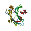
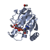
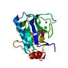
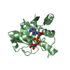
| Title | Getah virus macro domain in complex with ADPr in double open conformation | ||||||
 Components Components | Non-structural polyprotein | ||||||
 Keywords Keywords |  VIRAL PROTEIN / Macro domain / Getah virus VIRAL PROTEIN / Macro domain / Getah virus | ||||||
| Function / homology |  Function and homology information Function and homology informationhost cell filopodium / ADP-ribose 1''-phosphate phosphatase / mRNA methyltransferase activity / mRNA 5'-triphosphate monophosphatase activity / mRNA 5'-phosphatase / polynucleotide 5'-phosphatase activity / polynucleotide adenylyltransferase / poly(A) RNA polymerase activity / symbiont-mediated suppression of host mRNA transcription via inhibition of RNA polymerase II activity / 7-methylguanosine mRNA capping ...host cell filopodium / ADP-ribose 1''-phosphate phosphatase / mRNA methyltransferase activity / mRNA 5'-triphosphate monophosphatase activity / mRNA 5'-phosphatase / polynucleotide 5'-phosphatase activity / polynucleotide adenylyltransferase / poly(A) RNA polymerase activity / symbiont-mediated suppression of host mRNA transcription via inhibition of RNA polymerase II activity / 7-methylguanosine mRNA capping / cysteine-type peptidase activity / ribonucleoside triphosphate phosphatase activity / Transferases; Transferring one-carbon groups; Methyltransferases / host cell cytoplasmic vesicle membrane / cytoplasmic vesicle membrane / Transferases; Transferring phosphorus-containing groups; Nucleotidyltransferases / nucleoside-triphosphate phosphatase / Hydrolases; Acting on peptide bonds (peptidases); Cysteine endopeptidases / RNA helicase activity / RNA helicase / RNA-directed RNA polymerase / viral RNA genome replication / RNA-dependent RNA polymerase activity / DNA-templated transcription / host cell nucleus / GTP binding / host cell plasma membrane / ATP hydrolysis activity / proteolysis / RNA binding / ATP binding / membrane / metal ion binding / plasma membraneSimilarity search - Function | ||||||
| Biological species |  Getah virus Getah virus | ||||||
| Method | X-RAY DIFFRACTION / SYNCHROTRON / FOURIER SYNTHESIS / Resolution: 1.6 Å | ||||||
 Authors Authors | Sulzenbacher, G. / Ferreira Ramos, A.S. / Coutard, B. | ||||||
 Citation Citation | Journal: Sci Rep / Year: 2020 Title: Snapshots of ADP-ribose bound to Getah virus macro domain reveal an intriguing choreography. Authors: Ferreira-Ramos, A.S. / Sulzenbacher, G. / Canard, B. / Coutard, B. | ||||||
| History |
|
- Structure visualization
| Structure viewer | Molecule: MolmilJmol/JSmol |
|---|
- Downloads & links
-Download
| PDBx/mmCIF format | 6r0p.cif.gz | 85.6 KB | Display | PDBx/mmCIF format |
|---|---|---|---|---|
| PDB format | pdb6r0p.ent.gz | 63.7 KB | Display | PDB format |
| PDBx/mmJSON format | 6r0p.json.gz | Tree view | PDBx/mmJSON format | |
| Others |  Other downloads Other downloads |
-Validation report
| Arichive directory | https://data.pdbj.org/pub/pdb/validation_reports/r0/6r0pftp://data.pdbj.org/pub/pdb/validation_reports/r0/6r0p | HTTPS FTP |
|---|
-Related structure data
| Related structure data |  6qzuSC  6r0fC  6r0gC  6r0rC  6r0tC S: Starting model for refinement C: citing same article ( |
|---|---|
| Similar structure data |


-Links
 PDBj
PDBj

- Assembly
| Deposited unit | 
| ||||||||
|---|---|---|---|---|---|---|---|---|---|
| 1 | 
| ||||||||
| 2 | 
| ||||||||
| Unit cell |
|
-Components
| #1: Protein | Mass: 18120.512 Da / Num. of mol.: 2 Source method: isolated from a genetically manipulated source Details: synthetic gene / Source: (gene. exp.) Getah virus / Gene: nsP1234 / Plasmid: pDest14Details (production host): from ThermoFischer, Gateway cloning Production host:  Escherichia coli BL21(DE3) (bacteria) / Variant (production host): Rosetta pLysS / References: UniProt: A0A143SL92, UniProt: Q5Y389*PLUS Escherichia coli BL21(DE3) (bacteria) / Variant (production host): Rosetta pLysS / References: UniProt: A0A143SL92, UniProt: Q5Y389*PLUS#2: Chemical |   Mass: 561.332 Da / Num. of mol.: 2 / Source method: obtained synthetically / Formula: C15H25N5O14P2 Mass: 561.332 Da / Num. of mol.: 2 / Source method: obtained synthetically / Formula: C15H25N5O14P2#3: Chemical | ChemComp-ACT / | Acetate  Mass: 59.044 Da / Num. of mol.: 1 / Source method: obtained synthetically / Formula: C2H3O2 Mass: 59.044 Da / Num. of mol.: 1 / Source method: obtained synthetically / Formula: C2H3O2#4: Chemical | ChemComp-EDO / | Ethylene glycol  Mass: 62.068 Da / Num. of mol.: 1 / Source method: obtained synthetically / Formula: C2H6O2 Mass: 62.068 Da / Num. of mol.: 1 / Source method: obtained synthetically / Formula: C2H6O2#5: Water | ChemComp-HOH / | Water Mass: 18.015 Da / Num. of mol.: 279 / Source method: isolated from a natural source / Formula: H2O Mass: 18.015 Da / Num. of mol.: 279 / Source method: isolated from a natural source / Formula: H2O |
|---|
-Experimental details
-Experiment
| Experiment | Method: X-RAY DIFFRACTION / Number of used crystals: 1 |
|---|
- Sample preparation
| Crystal | Density Matthews: 2.29 Å3/Da / Density % sol: 46.3 % |
|---|---|
| Crystal grow | Temperature: 293 K / Method: vapor diffusion / pH: 6 Details: Imidazole-Malate pH 6.0, 32% PEG 4K, 3mM ADPr, 30mM Aspartic acid |
-Data collection
| Diffraction | Mean temperature: 100 K / Serial crystal experiment: N |
|---|---|
| Diffraction source | Source: SYNCHROTRON / Site: ESRF  / Beamline: MASSIF-3 / Wavelength: 0.9677 Å / Beamline: MASSIF-3 / Wavelength: 0.9677 Å |
| Detector | Type: DECTRIS EIGER X 4M / Detector: PIXEL / Date: Jan 31, 2018 |
| Radiation | Protocol: SINGLE WAVELENGTH / Monochromatic (M) / Laue (L): M / Scattering type: x-ray |
| Radiation wavelength | Wavelength: 0.9677 Å / Relative weight: 1 |
| Reflection | Resolution: 1.6→40.83 Å / Num. obs: 44233 / % possible obs: 98.9 % / Observed criterion σ(F): 0 / Observed criterion σ(I): 0 / Redundancy: 4 % / Biso Wilson estimate: 15.4 Å2 / CC1/2: 0.998 / Rmerge(I) obs: 0.053 / Rpim(I) all: 0.043 / Rrim(I) all: 0.069 / Rsym value: 0.053 / Net I/σ(I): 13.1 |
| Reflection shell | Resolution: 1.6→1.63 Å / Redundancy: 4.1 % / Rmerge(I) obs: 1.119 / Mean I/σ(I) obs: 1.2 / Num. unique obs: 2174 / CC1/2: 0.559 / Rpim(I) all: 0.915 / Rrim(I) all: 1.451 / Rsym value: 1.119 / % possible all: 99.5 |
- Processing
| Software |
| ||||||||||||||||||||||||||||||||||||||||||||||||||||||||||||||||||||||||||||||||||||||||||||||||||||||||||||||||||||||||||||||||||||||||||||||||||||||||||||||||||||||||||||||||||||||
|---|---|---|---|---|---|---|---|---|---|---|---|---|---|---|---|---|---|---|---|---|---|---|---|---|---|---|---|---|---|---|---|---|---|---|---|---|---|---|---|---|---|---|---|---|---|---|---|---|---|---|---|---|---|---|---|---|---|---|---|---|---|---|---|---|---|---|---|---|---|---|---|---|---|---|---|---|---|---|---|---|---|---|---|---|---|---|---|---|---|---|---|---|---|---|---|---|---|---|---|---|---|---|---|---|---|---|---|---|---|---|---|---|---|---|---|---|---|---|---|---|---|---|---|---|---|---|---|---|---|---|---|---|---|---|---|---|---|---|---|---|---|---|---|---|---|---|---|---|---|---|---|---|---|---|---|---|---|---|---|---|---|---|---|---|---|---|---|---|---|---|---|---|---|---|---|---|---|---|---|---|---|---|---|
| Refinement | Method to determine structure: FOURIER SYNTHESIS Starting model: 6QZU Resolution: 1.6→36.41 Å / Cor.coef. Fo:Fc: 0.972 / Cor.coef. Fo:Fc free: 0.962 / SU B: 1.841 / SU ML: 0.06 / Cross valid method: THROUGHOUT / ESU R: 0.086 / ESU R Free: 0.083 / Details: HYDROGENS HAVE BEEN ADDED IN THE RIDING POSITIONS
| ||||||||||||||||||||||||||||||||||||||||||||||||||||||||||||||||||||||||||||||||||||||||||||||||||||||||||||||||||||||||||||||||||||||||||||||||||||||||||||||||||||||||||||||||||||||
| Solvent computation | Ion probe radii: 0.7 Å / Shrinkage radii: 0.7 Å / VDW probe radii: 1.1 Å | ||||||||||||||||||||||||||||||||||||||||||||||||||||||||||||||||||||||||||||||||||||||||||||||||||||||||||||||||||||||||||||||||||||||||||||||||||||||||||||||||||||||||||||||||||||||
| Displacement parameters | Biso mean: 25.39 Å2
| ||||||||||||||||||||||||||||||||||||||||||||||||||||||||||||||||||||||||||||||||||||||||||||||||||||||||||||||||||||||||||||||||||||||||||||||||||||||||||||||||||||||||||||||||||||||
| Refinement step | Cycle: 1 / Resolution: 1.6→36.41 Å
| ||||||||||||||||||||||||||||||||||||||||||||||||||||||||||||||||||||||||||||||||||||||||||||||||||||||||||||||||||||||||||||||||||||||||||||||||||||||||||||||||||||||||||||||||||||||
| Refine LS restraints |
|