 Movie
Movie Controller

[English] 日本語
 Yorodumi
- PDB-2xii: CRYSTAL STRUCTURE OF AN ALPHA-L-FUCOSIDASE GH29 FROM BACTEROIDES ... -
+ Open data

- Basic information
| Entry | Database: PDB / ID: 2xii | ||||||
|---|---|---|---|---|---|---|---|
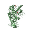
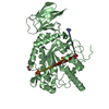
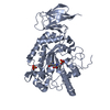
| Title | CRYSTAL STRUCTURE OF AN ALPHA-L-FUCOSIDASE GH29 FROM BACTEROIDES THETAIOTAOMICRON IN COMPLEX WITH AN EXTENDED 9-FLUORENONE IMINOSUGAR INHIBITOR | ||||||
 Components Components | ALPHA-L-FUCOSIDASE | ||||||
 Keywords Keywords | HYDROLASE / FUCOSE | ||||||
| Function / homology |  Function and homology informationalpha-L-fucosidase activity / fucose metabolic process / glycoside catabolic process / lysosome Function and homology informationalpha-L-fucosidase activity / fucose metabolic process / glycoside catabolic process / lysosomeSimilarity search - Function | ||||||
| Biological species |  BACTEROIDES THETAIOTAOMICRON (bacteria) BACTEROIDES THETAIOTAOMICRON (bacteria) | ||||||
| Method | X-RAY DIFFRACTION / SYNCHROTRON / MOLECULAR REPLACEMENT / Resolution: 1.8 Å | ||||||
 Authors Authors | Lammerts van Bueren, A. / Popat, S.D. / Lin, C.H. / Davies, G.J. | ||||||
 Citation Citation | Journal: Chembiochem / Year: 2010 Title: Structural and Thermodynamic Analyses of Alpha-L-Fucosidase Inhibitors. Authors: Lammerts Van Bueren, A. / Popat, S.D. / Lin, C.H. / Davies, G.J. | ||||||
| History |
|
- Structure visualization
| Structure viewer | Molecule: MolmilJmol/JSmol |
|---|
- Downloads & links
-Download
| PDBx/mmCIF format | 2xii.cif.gz | 204.9 KB | Display | PDBx/mmCIF format |
|---|---|---|---|---|
| PDB format | pdb2xii.ent.gz | 162.6 KB | Display | PDB format |
| PDBx/mmJSON format | 2xii.json.gz | Tree view | PDBx/mmJSON format | |
| Others |  Other downloads Other downloads |
-Validation report
| Arichive directory | https://data.pdbj.org/pub/pdb/validation_reports/xi/2xiiftp://data.pdbj.org/pub/pdb/validation_reports/xi/2xii | HTTPS FTP |
|---|
-Related structure data
| Related structure data |  2xibC  2wvtS S: Starting model for refinement C: citing same article ( |
|---|---|
| Similar structure data |


-Links
 PDBj
PDBj

- Assembly
| Deposited unit | 
| ||||||||
|---|---|---|---|---|---|---|---|---|---|
| 1 | 
| ||||||||
| 2 | 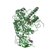
| ||||||||
| Unit cell |
|
-Components
-Protein , 1 types, 2 molecules AB
| #1: Protein | / FUCOSIDASE / GLYCOSIDE HYDROLASE / ALPHA-L-FUCOSIDE FUCOHYDROLASE Mass: 52410.367 Da / Num. of mol.: 2 / Fragment: CATALYTIC DOMAIN, 32-484 Source method: isolated from a genetically manipulated source Source: (gene. exp.) BACTEROIDES THETAIOTAOMICRON (bacteria)Production host: ESCHERICHIA COLI (E. coli) / Strain (production host): BL21(DE3) / References: UniProt: Q8A3I4, alpha-L-fucosidase |
|---|
-Non-polymers , 5 types, 705 molecules 


| #2: Chemical | Tyrosine Type: L-peptide linking / Mass: 181.189 Da / Num. of mol.: 2 / Source method: obtained synthetically / Formula: C9H11NO3 Type: L-peptide linking / Mass: 181.189 Da / Num. of mol.: 2 / Source method: obtained synthetically / Formula: C9H11NO3#3: Chemical |  Mass: 382.410 Da / Num. of mol.: 2 / Source method: obtained synthetically / Formula: C21H22N2O5 Mass: 382.410 Da / Num. of mol.: 2 / Source method: obtained synthetically / Formula: C21H22N2O5#4: Chemical | ChemComp-SO4 / Sulfate Mass: 96.063 Da / Num. of mol.: 4 / Source method: obtained synthetically / Formula: SO4 Mass: 96.063 Da / Num. of mol.: 4 / Source method: obtained synthetically / Formula: SO4#5: Chemical | ChemComp-GOL / | Glycerol Mass: 92.094 Da / Num. of mol.: 1 / Source method: obtained synthetically / Formula: C3H8O3 Mass: 92.094 Da / Num. of mol.: 1 / Source method: obtained synthetically / Formula: C3H8O3#6: Water | ChemComp-HOH / | WaterMass: 18.015 Da / Num. of mol.: 696 / Source method: isolated from a natural source / Formula: H2O |
|---|
-Experimental details
-Experiment
| Experiment | Method: X-RAY DIFFRACTION / Number of used crystals: 1 |
|---|
- Sample preparation
| Crystal | Density Matthews: 2.42 Å3/Da / Density % sol: 49.09 % / Description: NONE |
|---|---|
| Crystal grow | Details: 20% PEG-6000, 0.15M AMMONIUM SULFATE, 0.1M IMIDAZOLE PH 7.0 |
-Data collection
| Diffraction | Mean temperature: 100 K |
|---|---|
| Diffraction source | Source: SYNCHROTRON / Site: ESRF  / Beamline: ID29 / Wavelength: 0.98 / Beamline: ID29 / Wavelength: 0.98 |
| Detector | Type: ADSC QUANTUM 315r / Detector: CCD / Date: Nov 30, 2009 |
| Radiation | Protocol: SINGLE WAVELENGTH / Monochromatic (M) / Laue (L): M / Scattering type: x-ray |
| Radiation wavelength | Wavelength: 0.98 Å / Relative weight: 1 |
| Reflection | Resolution: 1.8→45 Å / Num. obs: 92257 / % possible obs: 99.9 % / Observed criterion σ(I): 2 / Redundancy: 3.6 % / Rmerge(I) obs: 0.08 / Net I/σ(I): 13.2 |
| Reflection shell | Resolution: 1.8→1.9 Å / Redundancy: 3.7 % / Rmerge(I) obs: 0.67 / Mean I/σ(I) obs: 3.7 / % possible all: 100 |
- Processing
| Software |
| ||||||||||||||||||||||||||||||||||||||||||||||||||||||||||||||||||||||||||||||||||||||||||||||||||||||||||||||||||||||||||||||||||||||||||||||||||||||||||||||||||||||||||||||||||||||
|---|---|---|---|---|---|---|---|---|---|---|---|---|---|---|---|---|---|---|---|---|---|---|---|---|---|---|---|---|---|---|---|---|---|---|---|---|---|---|---|---|---|---|---|---|---|---|---|---|---|---|---|---|---|---|---|---|---|---|---|---|---|---|---|---|---|---|---|---|---|---|---|---|---|---|---|---|---|---|---|---|---|---|---|---|---|---|---|---|---|---|---|---|---|---|---|---|---|---|---|---|---|---|---|---|---|---|---|---|---|---|---|---|---|---|---|---|---|---|---|---|---|---|---|---|---|---|---|---|---|---|---|---|---|---|---|---|---|---|---|---|---|---|---|---|---|---|---|---|---|---|---|---|---|---|---|---|---|---|---|---|---|---|---|---|---|---|---|---|---|---|---|---|---|---|---|---|---|---|---|---|---|---|---|
| Refinement | Method to determine structure: MOLECULAR REPLACEMENT Starting model: PDB ENTRY 2WVT Resolution: 1.8→45.15 Å / Cor.coef. Fo:Fc: 0.96 / Cor.coef. Fo:Fc free: 0.943 / SU B: 3.079 / SU ML: 0.094 / Cross valid method: THROUGHOUT / ESU R: 0.13 / ESU R Free: 0.123 / Stereochemistry target values: MAXIMUM LIKELIHOOD Details: HYDROGENS HAVE BEEN ADDED IN THE RIDING POSITIONS. U VALUES REFINED INDIVIDUALLY
| ||||||||||||||||||||||||||||||||||||||||||||||||||||||||||||||||||||||||||||||||||||||||||||||||||||||||||||||||||||||||||||||||||||||||||||||||||||||||||||||||||||||||||||||||||||||
| Solvent computation | Ion probe radii: 0.8 Å / Shrinkage radii: 0.8 Å / VDW probe radii: 1.4 Å / Solvent model: MASK | ||||||||||||||||||||||||||||||||||||||||||||||||||||||||||||||||||||||||||||||||||||||||||||||||||||||||||||||||||||||||||||||||||||||||||||||||||||||||||||||||||||||||||||||||||||||
| Displacement parameters | Biso mean: 20.537 Å2
| ||||||||||||||||||||||||||||||||||||||||||||||||||||||||||||||||||||||||||||||||||||||||||||||||||||||||||||||||||||||||||||||||||||||||||||||||||||||||||||||||||||||||||||||||||||||
| Refinement step | Cycle: LAST / Resolution: 1.8→45.15 Å
| ||||||||||||||||||||||||||||||||||||||||||||||||||||||||||||||||||||||||||||||||||||||||||||||||||||||||||||||||||||||||||||||||||||||||||||||||||||||||||||||||||||||||||||||||||||||
| Refine LS restraints |
|