 Movie
Movie Controller

[English] 日本語
 Yorodumi
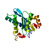
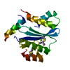
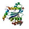
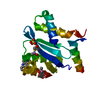
- PDB-2xgv: Structure of the N-terminal domain of capsid protein from Rabbit ... -
+ Open data

- Basic information
| Entry | Database: PDB / ID: 2xgv | ||||||
|---|---|---|---|---|---|---|---|
| Title | Structure of the N-terminal domain of capsid protein from Rabbit Endogenous Lentivirus (RELIK) | ||||||
 Components Components | PSIV CAPSID N-TERMINAL DOMAIN | ||||||
 Keywords Keywords |  VIRAL PROTEIN / RETROVIRAL CAPSID VIRAL PROTEIN / RETROVIRAL CAPSID | ||||||
| Function / homology | Human Immunodeficiency Virus Type 1 Capsid Protein / Human Immunodeficiency Virus Type 1 Capsid Protein / Orthogonal Bundle / Mainly Alpha Function and homology information Function and homology information | ||||||
| Biological species |  MICROCEBUS MURINUS (gray mouse lemur) MICROCEBUS MURINUS (gray mouse lemur) | ||||||
| Method | X-RAY DIFFRACTION / MOLECULAR REPLACEMENT / Resolution: 2 Å | ||||||
 Authors Authors | Goldstone, D.C. / Robertson, L.E. / Haire, L.F. / Stoye, J.P. / Taylor, I.A. | ||||||
 Citation Citation | Journal: Cell Host Microbe / Year: 2010 Title: Structural and Functional Analysis of Prehistoric Lentiviruses Uncovers an Ancient Molecular Interface. Authors: Goldstone, D.C. / Yap, M.W. / Robertson, L.E. / Haire, L.F. / Taylor, W.R. / Katzourakis, A. / Stoye, J.P. / Taylor, I.A. | ||||||
| History |
|
- Structure visualization
| Structure viewer | Molecule: MolmilJmol/JSmol |
|---|
- Downloads & links
-Download
| PDBx/mmCIF format | 2xgv.cif.gz | 66.4 KB | Display | PDBx/mmCIF format |
|---|---|---|---|---|
| PDB format | pdb2xgv.ent.gz | 49.5 KB | Display | PDB format |
| PDBx/mmJSON format | 2xgv.json.gz | Tree view | PDBx/mmJSON format | |
| Others |  Other downloads Other downloads |
-Validation report
| Arichive directory | https://data.pdbj.org/pub/pdb/validation_reports/xg/2xgvftp://data.pdbj.org/pub/pdb/validation_reports/xg/2xgv | HTTPS FTP |
|---|
-Related structure data


-Links
 PDBj
PDBj- Assembly
| Deposited unit | 
| ||||||||
|---|---|---|---|---|---|---|---|---|---|
| 1 |
| ||||||||
| Unit cell |
| ||||||||
| Components on special symmetry positions |
|
-Components
| #1: Protein | Mass: 15993.886 Da / Num. of mol.: 1 Source method: isolated from a genetically manipulated source Source: (gene. exp.) MICROCEBUS MURINUS (gray mouse lemur) / Production host:  ESCHERICHIA COLI (E. coli) ESCHERICHIA COLI (E. coli) |
|---|---|
| #2: Water | ChemComp-HOH / Water Mass: 18.015 Da / Num. of mol.: 110 / Source method: isolated from a natural source / Formula: H2O Mass: 18.015 Da / Num. of mol.: 110 / Source method: isolated from a natural source / Formula: H2O |
-Experimental details
-Experiment
| Experiment | Method: X-RAY DIFFRACTION |
|---|
- Sample preparation
| Crystal | Density Matthews: 2.07 Å3/Da / Density % sol: 40.67 % / Description: NONE |
|---|---|
| Crystal grow | pH: 4.6 / Details: 24% PEG6000, 0.1M NA-ACETATE PH4.6. |
-Data collection
| Diffraction | Mean temperature: 100 K |
|---|---|
| Diffraction source | Source: ROTATING ANODE / Wavelength: 1.5418 |
| Detector | Type: RIGAKU-MSC / Detector: AREA DETECTOR |
| Radiation | Protocol: SINGLE WAVELENGTH / Monochromatic (M) / Laue (L): M / Scattering type: x-ray |
| Radiation wavelength | Wavelength: 1.5418 Å / Relative weight: 1 |
| Reflection | Resolution: 2→25 Å / Num. obs: 8728 / % possible obs: 98.7 % / Observed criterion σ(I): 2 / Redundancy: 3.5 % / Biso Wilson estimate: 23.3 Å2 / Rmerge(I) obs: 0.06 / Net I/σ(I): 11.6 |
| Reflection shell | Resolution: 2→2.11 Å / Redundancy: 3.4 % / Rmerge(I) obs: 0.24 / Mean I/σ(I) obs: 4.3 / % possible all: 96.9 |
- Processing
| Software |
| |||||||||||||||||||||||||||||||||||||||||||||||||||||||||||||||||||||||||||||||||||||||||||||||||||||||||||||||||||||||||||||||||||||||||||||||||||||||||||||||||||||||||||||||||||||||||||||||||||||||||||||||||||||||||||||||||
|---|---|---|---|---|---|---|---|---|---|---|---|---|---|---|---|---|---|---|---|---|---|---|---|---|---|---|---|---|---|---|---|---|---|---|---|---|---|---|---|---|---|---|---|---|---|---|---|---|---|---|---|---|---|---|---|---|---|---|---|---|---|---|---|---|---|---|---|---|---|---|---|---|---|---|---|---|---|---|---|---|---|---|---|---|---|---|---|---|---|---|---|---|---|---|---|---|---|---|---|---|---|---|---|---|---|---|---|---|---|---|---|---|---|---|---|---|---|---|---|---|---|---|---|---|---|---|---|---|---|---|---|---|---|---|---|---|---|---|---|---|---|---|---|---|---|---|---|---|---|---|---|---|---|---|---|---|---|---|---|---|---|---|---|---|---|---|---|---|---|---|---|---|---|---|---|---|---|---|---|---|---|---|---|---|---|---|---|---|---|---|---|---|---|---|---|---|---|---|---|---|---|---|---|---|---|---|---|---|---|---|---|---|---|---|---|---|---|---|---|---|---|---|---|---|---|---|
| Refinement | Method to determine structure: MOLECULAR REPLACEMENT / Resolution: 2→24.529 Å / SU ML: 0.31 / σ(F): 1.43 / Phase error: 22.9 / Stereochemistry target values: ML
| |||||||||||||||||||||||||||||||||||||||||||||||||||||||||||||||||||||||||||||||||||||||||||||||||||||||||||||||||||||||||||||||||||||||||||||||||||||||||||||||||||||||||||||||||||||||||||||||||||||||||||||||||||||||||||||||||
| Solvent computation | Shrinkage radii: 0.9 Å / VDW probe radii: 1.11 Å / Solvent model: FLAT BULK SOLVENT MODEL / Bsol: 40.914 Å2 / ksol: 0.339 e/Å3 | |||||||||||||||||||||||||||||||||||||||||||||||||||||||||||||||||||||||||||||||||||||||||||||||||||||||||||||||||||||||||||||||||||||||||||||||||||||||||||||||||||||||||||||||||||||||||||||||||||||||||||||||||||||||||||||||||
| Displacement parameters | Biso mean: 26.7 Å2
| |||||||||||||||||||||||||||||||||||||||||||||||||||||||||||||||||||||||||||||||||||||||||||||||||||||||||||||||||||||||||||||||||||||||||||||||||||||||||||||||||||||||||||||||||||||||||||||||||||||||||||||||||||||||||||||||||
| Refinement step | Cycle: LAST / Resolution: 2→24.529 Å
| |||||||||||||||||||||||||||||||||||||||||||||||||||||||||||||||||||||||||||||||||||||||||||||||||||||||||||||||||||||||||||||||||||||||||||||||||||||||||||||||||||||||||||||||||||||||||||||||||||||||||||||||||||||||||||||||||
| Refine LS restraints |
| |||||||||||||||||||||||||||||||||||||||||||||||||||||||||||||||||||||||||||||||||||||||||||||||||||||||||||||||||||||||||||||||||||||||||||||||||||||||||||||||||||||||||||||||||||||||||||||||||||||||||||||||||||||||||||||||||
| LS refinement shell |
| |||||||||||||||||||||||||||||||||||||||||||||||||||||||||||||||||||||||||||||||||||||||||||||||||||||||||||||||||||||||||||||||||||||||||||||||||||||||||||||||||||||||||||||||||||||||||||||||||||||||||||||||||||||||||||||||||
| Refinement TLS params. | Method: refined / Refine-ID: X-RAY DIFFRACTION
| |||||||||||||||||||||||||||||||||||||||||||||||||||||||||||||||||||||||||||||||||||||||||||||||||||||||||||||||||||||||||||||||||||||||||||||||||||||||||||||||||||||||||||||||||||||||||||||||||||||||||||||||||||||||||||||||||
| Refinement TLS group |
|
