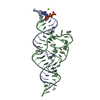
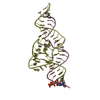
 Movie
Movie Controller


+ Open data

- Basic information
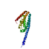
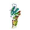
| Entry | Database: PDB / ID: 2wvi | ||||||
|---|---|---|---|---|---|---|---|
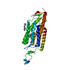
| Title | Crystal Structure of the N-terminal Domain of BubR1 | ||||||
 Components Components | MITOTIC CHECKPOINT SERINE/THREONINE-PROTEIN KINASE BUB1 BETA | ||||||
 Keywords Keywords |  TRANSFERASE / TUMOR SUPPRESSOR / MITOTIC CHECKPOINT / TPR / KINASE / APOPTOSIS / SERINE/THREONINE-PROTEIN KINASE / CELL DIVISION / CELL CYCLE / KINETOCHORE / CYTOSKELETON TRANSFERASE / TUMOR SUPPRESSOR / MITOTIC CHECKPOINT / TPR / KINASE / APOPTOSIS / SERINE/THREONINE-PROTEIN KINASE / CELL DIVISION / CELL CYCLE / KINETOCHORE / CYTOSKELETON | ||||||
| Function / homology |  Function and homology informationmitotic checkpoint complex / meiotic sister chromatid cohesion, centromeric / Inactivation of APC/C via direct inhibition of the APC/C complex / APC/C:Cdc20 mediated degradation of mitotic proteins / anaphase-promoting complex / metaphase/anaphase transition of mitotic cell cycle / outer kinetochore / protein localization to chromosome, centromeric region / mitotic spindle assembly checkpoint signaling / Amplification of signal from unattached kinetochores via a MAD2 inhibitory signal ...mitotic checkpoint complex / meiotic sister chromatid cohesion, centromeric / Inactivation of APC/C via direct inhibition of the APC/C complex / APC/C:Cdc20 mediated degradation of mitotic proteins / anaphase-promoting complex / metaphase/anaphase transition of mitotic cell cycle / outer kinetochore / protein localization to chromosome, centromeric region / mitotic spindle assembly checkpoint signaling / Amplification of signal from unattached kinetochores via a MAD2 inhibitory signal / Mitotic Prometaphase / EML4 and NUDC in mitotic spindle formation / Resolution of Sister Chromatid Cohesion / APC-Cdc20 mediated degradation of Nek2A / RHO GTPases Activate Formins / Cdc20:Phospho-APC/C mediated degradation of Cyclin A / kinetochore / spindle / Separation of Sister Chromatids / non-specific serine/threonine protein kinase / protein kinase activity / cell division / phosphorylation / protein serine kinase activity / protein serine/threonine kinase activity / centrosome / apoptotic process / perinuclear region of cytoplasm / ATP binding / nucleus / cytosol / cytoplasm Function and homology informationmitotic checkpoint complex / meiotic sister chromatid cohesion, centromeric / Inactivation of APC/C via direct inhibition of the APC/C complex / APC/C:Cdc20 mediated degradation of mitotic proteins / anaphase-promoting complex / metaphase/anaphase transition of mitotic cell cycle / outer kinetochore / protein localization to chromosome, centromeric region / mitotic spindle assembly checkpoint signaling / Amplification of signal from unattached kinetochores via a MAD2 inhibitory signal ...mitotic checkpoint complex / meiotic sister chromatid cohesion, centromeric / Inactivation of APC/C via direct inhibition of the APC/C complex / APC/C:Cdc20 mediated degradation of mitotic proteins / anaphase-promoting complex / metaphase/anaphase transition of mitotic cell cycle / outer kinetochore / protein localization to chromosome, centromeric region / mitotic spindle assembly checkpoint signaling / Amplification of signal from unattached kinetochores via a MAD2 inhibitory signal / Mitotic Prometaphase / EML4 and NUDC in mitotic spindle formation / Resolution of Sister Chromatid Cohesion / APC-Cdc20 mediated degradation of Nek2A / RHO GTPases Activate Formins / Cdc20:Phospho-APC/C mediated degradation of Cyclin A / kinetochore / spindle / Separation of Sister Chromatids / non-specific serine/threonine protein kinase / protein kinase activity / cell division / phosphorylation / protein serine kinase activity / protein serine/threonine kinase activity / centrosome / apoptotic process / perinuclear region of cytoplasm / ATP binding / nucleus / cytosol / cytoplasmSimilarity search - Function | ||||||
| Biological species |  HOMO SAPIENS (human) HOMO SAPIENS (human) | ||||||
| Method | X-RAY DIFFRACTION / SYNCHROTRON / SAD / Resolution: 1.8 Å | ||||||
 Authors Authors | D'Arcy, S. / Davies, O.R. / Blundell, T.L. / Bolanos-Garcia, V.M. | ||||||
 Citation Citation | Journal: J.Biol.Chem. / Year: 2010 Title: Defining the Molecular Basis of Bubr1 Kinetochore Interactions and Anaphase-Promoting Complex/Cyclosome (Apc/C)-Cdc20 Inhibition Authors: D'Arcy, S. / Davies, O.R. / Blundell, T.L. / Bolanos-Garcia, V.M. | ||||||
| History |
|
- Structure visualization
| Structure viewer | Molecule: MolmilJmol/JSmol |
|---|
- Downloads & links
-Download
| PDBx/mmCIF format | 2wvi.cif.gz | 51.8 KB | Display | PDBx/mmCIF format |
|---|---|---|---|---|
| PDB format | pdb2wvi.ent.gz | 37 KB | Display | PDB format |
| PDBx/mmJSON format | 2wvi.json.gz | Tree view | PDBx/mmJSON format | |
| Others |  Other downloads Other downloads |
-Validation report
| Arichive directory | https://data.pdbj.org/pub/pdb/validation_reports/wv/2wviftp://data.pdbj.org/pub/pdb/validation_reports/wv/2wvi | HTTPS FTP |
|---|
-Related structure data
| Similar structure data |
|---|


-Links
 PDBj
PDBj


- Assembly
| Deposited unit | 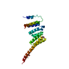
| ||||||||
|---|---|---|---|---|---|---|---|---|---|
| 1 |
| ||||||||
| Unit cell |
|
-Components
| #1: Protein | Mass: 19597.682 Da / Num. of mol.: 1 / Fragment: N-TERMINAL TPR DOMAIN, RESIDUES 57-220 Source method: isolated from a genetically manipulated source Source: (gene. exp.) HOMO SAPIENS (human) / Plasmid: PHAT4 / Production host:  ESCHERICHIA COLI (E. coli) / Strain (production host): BL21(DE3) ESCHERICHIA COLI (E. coli) / Strain (production host): BL21(DE3)References: UniProt: O60566, non-specific serine/threonine protein kinase | ||||||||
|---|---|---|---|---|---|---|---|---|---|
| #2: Chemical | Praseodymium  Mass: 140.908 Da / Num. of mol.: 2 / Source method: obtained synthetically / Formula: Pr Mass: 140.908 Da / Num. of mol.: 2 / Source method: obtained synthetically / Formula: Pr#3: Chemical | ChemComp-ACT / | Acetate  Mass: 59.044 Da / Num. of mol.: 1 / Source method: obtained synthetically / Formula: C2H3O2 Mass: 59.044 Da / Num. of mol.: 1 / Source method: obtained synthetically / Formula: C2H3O2#4: Chemical | Ethylene glycol  Mass: 62.068 Da / Num. of mol.: 2 / Source method: obtained synthetically / Formula: C2H6O2 Mass: 62.068 Da / Num. of mol.: 2 / Source method: obtained synthetically / Formula: C2H6O2#5: Water | ChemComp-HOH / | Water Mass: 18.015 Da / Num. of mol.: 117 / Source method: isolated from a natural source / Formula: H2O Mass: 18.015 Da / Num. of mol.: 117 / Source method: isolated from a natural source / Formula: H2ONonpolymer details | ETHANE-1,2-DIOL (EDO): USED AS CRYOPROTECTANT PRASEODYMIUM ION (PR): ESSENTIAL FOR HIGH RESOLUTION ...ETHANE-1,2-DIOL (EDO): USED AS CRYOPROTEC | |
-Experimental details
-Experiment
| Experiment | Method: X-RAY DIFFRACTION / Number of used crystals: 2 |
|---|
- Sample preparation
| Crystal | Density Matthews: 2.47 Å3/Da / Density % sol: 49 % / Description: THIS DATA SET WAS USED IN REFINEMENTS. |
|---|---|
| Crystal grow | pH: 8 Details: 22.5% (W/V) PEG 8000, 0.17 M TRIS PH 8.0, 0.01 M PRASEODYMIUM(III) ACETATE |
-Data collection
| Diffraction | Mean temperature: 100 K |
|---|---|
| Diffraction source | Source: SYNCHROTRON / Site: Diamond  / Beamline: I03 / Wavelength: 1.06 / Beamline: I03 / Wavelength: 1.06 |
| Detector | Type: ADSC CCD / Detector: CCD / Date: Oct 12, 2007 Details: KIRKPATRICK BAEZ BIMORPH MIRROR PAIR FOR HORIZONTAL AND VERTICAL FOCUSSING |
| Radiation | Monochromator: SI (111) DOUBLE CRYSTAL MONOCHROMATOR / Protocol: SINGLE WAVELENGTH / Monochromatic (M) / Laue (L): M / Scattering type: x-ray |
| Radiation wavelength | Wavelength: 1.06 Å / Relative weight: 1 |
| Reflection | Resolution: 1.8→46.62 Å / Num. obs: 19685 / % possible obs: 99.9 % / Observed criterion σ(I): 2 / Redundancy: 15.6 % / Biso Wilson estimate: 22 Å2 / Rmerge(I) obs: 0.11 / Net I/σ(I): 21 |
| Reflection shell | Resolution: 1.8→1.9 Å / Redundancy: 15.8 % / Rmerge(I) obs: 0.51 / Mean I/σ(I) obs: 4 / % possible all: 100 |
- Processing
| Software |
| ||||||||||||||||||||||||||||||||||||||||||||||||||||||||||||||||||||||||||||||||||||||||||||||||||||||||||||||||||||||||||||||||||||||||||||||||||||||||||||||||||||||||||||||||||||||
|---|---|---|---|---|---|---|---|---|---|---|---|---|---|---|---|---|---|---|---|---|---|---|---|---|---|---|---|---|---|---|---|---|---|---|---|---|---|---|---|---|---|---|---|---|---|---|---|---|---|---|---|---|---|---|---|---|---|---|---|---|---|---|---|---|---|---|---|---|---|---|---|---|---|---|---|---|---|---|---|---|---|---|---|---|---|---|---|---|---|---|---|---|---|---|---|---|---|---|---|---|---|---|---|---|---|---|---|---|---|---|---|---|---|---|---|---|---|---|---|---|---|---|---|---|---|---|---|---|---|---|---|---|---|---|---|---|---|---|---|---|---|---|---|---|---|---|---|---|---|---|---|---|---|---|---|---|---|---|---|---|---|---|---|---|---|---|---|---|---|---|---|---|---|---|---|---|---|---|---|---|---|---|---|
| Refinement | Method to determine structure: SAD Starting model: NONE Resolution: 1.8→54.39 Å / Cor.coef. Fo:Fc: 0.943 / Cor.coef. Fo:Fc free: 0.93 / SU B: 3.444 / SU ML: 0.106 / Cross valid method: THROUGHOUT / ESU R: 0.139 / ESU R Free: 0.135 / Stereochemistry target values: MAXIMUM LIKELIHOOD Details: HYDROGENS HAVE BEEN ADDED IN THE RIDING POSITIONS. DENSITY WAS NOT OBSERVED FOR RESIDUES 92-95. ONE ROUND OF TLS WAS CONDUCTED DURING REFINEMENT. TWO GROUPS WERE USED; RESIDUES 57-114 AND 115-220.
| ||||||||||||||||||||||||||||||||||||||||||||||||||||||||||||||||||||||||||||||||||||||||||||||||||||||||||||||||||||||||||||||||||||||||||||||||||||||||||||||||||||||||||||||||||||||
| Solvent computation | Ion probe radii: 0.8 Å / Shrinkage radii: 0.8 Å / VDW probe radii: 1.4 Å / Solvent model: MASK | ||||||||||||||||||||||||||||||||||||||||||||||||||||||||||||||||||||||||||||||||||||||||||||||||||||||||||||||||||||||||||||||||||||||||||||||||||||||||||||||||||||||||||||||||||||||
| Displacement parameters | Biso mean: 37.105 Å2
| ||||||||||||||||||||||||||||||||||||||||||||||||||||||||||||||||||||||||||||||||||||||||||||||||||||||||||||||||||||||||||||||||||||||||||||||||||||||||||||||||||||||||||||||||||||||
| Refinement step | Cycle: LAST / Resolution: 1.8→54.39 Å
| ||||||||||||||||||||||||||||||||||||||||||||||||||||||||||||||||||||||||||||||||||||||||||||||||||||||||||||||||||||||||||||||||||||||||||||||||||||||||||||||||||||||||||||||||||||||
| Refine LS restraints |
|