 Movie
Movie Controller


+ Open data

- Basic information
| Entry | Database: PDB / ID: 2wtl | ||||||
|---|---|---|---|---|---|---|---|
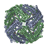
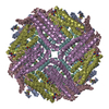
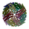
| Title | Crystal structure of BfrA from M. tuberculosis | ||||||
 Components Components | BACTERIOFERRITIN | ||||||
 Keywords Keywords | METAL BINDING PROTEIN / BACTERIOFERRITIN A / HEME / IRON / BILIVERDIN / IRON STORAGE / METAL-BINDING | ||||||
| Function / homology |  Function and homology information Function and homology informationMtb iron assimilation by chelation / response to iron ion / ferroxidase / ferroxidase activity / intracellular sequestering of iron ion / ferric iron binding / iron ion transport / intracellular iron ion homeostasis / iron ion binding / heme binding ...Mtb iron assimilation by chelation / response to iron ion / ferroxidase / ferroxidase activity / intracellular sequestering of iron ion / ferric iron binding / iron ion transport / intracellular iron ion homeostasis / iron ion binding / heme binding / extracellular region / plasma membrane / cytosolSimilarity search - Function | ||||||
| Biological species |   MYCOBACTERIUM TUBERCULOSIS (bacteria) MYCOBACTERIUM TUBERCULOSIS (bacteria) | ||||||
| Method | X-RAY DIFFRACTION / SYNCHROTRON / MOLECULAR REPLACEMENT / Resolution: 2.59 Å | ||||||
 Authors Authors | Gupta, V. / Gupta, R.K. / Khare, G. / Salunke, D.M. / Tyagi, A.K. | ||||||
 Citation Citation | Journal: Plos One / Year: 2009 Title: Crystal Structure of Bfra from Mycobacterium Tuberculosis:Incorporation of Selenomethionine Results in Cleavage and Demetallation of Haem Authors: Gupta, V. / Gupta, R.K. / Khare, G. / Salunke, D.M. / Tyagi, A.K. | ||||||
| History |
|
- Structure visualization
| Structure viewer | Molecule: MolmilJmol/JSmol |
|---|
- Downloads & links
-Download
| PDBx/mmCIF format | 2wtl.cif.gz | 205.8 KB | Display | PDBx/mmCIF format |
|---|---|---|---|---|
| PDB format | pdb2wtl.ent.gz | 166.9 KB | Display | PDB format |
| PDBx/mmJSON format | 2wtl.json.gz | Tree view | PDBx/mmJSON format | |
| Others |  Other downloads Other downloads |
-Validation report
| Arichive directory | https://data.pdbj.org/pub/pdb/validation_reports/wt/2wtlftp://data.pdbj.org/pub/pdb/validation_reports/wt/2wtl | HTTPS FTP |
|---|
-Related structure data
| Related structure data | 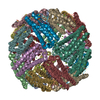 3bknS S: Starting model for refinement |
|---|---|
| Similar structure data |


-Links
 PDBj
PDBj


- Assembly
| Deposited unit | 
| ||||||||||||||||||||||||
|---|---|---|---|---|---|---|---|---|---|---|---|---|---|---|---|---|---|---|---|---|---|---|---|---|---|
| 1 | 
| ||||||||||||||||||||||||
| Unit cell |
| ||||||||||||||||||||||||
| Components on special symmetry positions |
| ||||||||||||||||||||||||
| Noncrystallographic symmetry (NCS) | NCS oper:
|
-Components
| #1: Protein | / BFR Mass: 20096.705 Da / Num. of mol.: 6 Source method: isolated from a genetically manipulated source Details: THIS IS A SELENOMETHIONYL ANALOG OF BFRA FROM M. TUBERCULOSIS (RV1876) WITH MSE31, MSE52, MSE107 AND MSE141 INSTEAD OF M31, M52, M107 AND M141 Source: (gene. exp.) MYCOBACTERIUM TUBERCULOSIS (bacteria) / Strain: H37RV / Description: LABORATORY STRAIN MYCOBACTERIUM TUBERCULOSIS / Plasmid: PET21C-BFRA / Production host: ESCHERICHIA COLI (E. coli) / Strain (production host): BL21(DE3) / References: UniProt: P63697, UniProt: P9WPQ9*PLUS#2: Chemical | ChemComp-FE / Iron  Mass: 55.845 Da / Num. of mol.: 12 / Source method: obtained synthetically / Formula: Fe Mass: 55.845 Da / Num. of mol.: 12 / Source method: obtained synthetically / Formula: Fe#3: Chemical |   Num. of mol.: 3 / Source method: obtained synthetically Num. of mol.: 3 / Source method: obtained synthetically#4: Chemical | Num. of mol.: 3 / Source method: obtained synthetically #5: Water | ChemComp-HOH / | Water Mass: 18.015 Da / Num. of mol.: 170 / Source method: isolated from a natural source / Formula: H2O Mass: 18.015 Da / Num. of mol.: 170 / Source method: isolated from a natural source / Formula: H2ONonpolymer details | UNKNOWN ATOM OR ION (UNX): UNKNOWN ION AT 4-FOLD AXIS PLACED WITH 0.25 OCCUPANCY UNKNOWN (UNL): ...UNKNOWN ATOM OR ION (UNX): UNKNOWN ION AT 4-FOLD AXIS PLACED WITH 0.25 OCCUPANCY UNKNOWN (UNL): POSSIBLE BILIVERDIN | |
|---|
-Experimental details
-Experiment
| Experiment | Method: X-RAY DIFFRACTION / Number of used crystals: 4 |
|---|
- Sample preparation
| Crystal | Density Matthews: 3.06 Å3/Da / Density % sol: 59.8 % / Description: NONE |
|---|---|
| Crystal grow | pH: 8 Details: PROTEIN (9MG/ML) WAS CRYSTALLIZED WITH 1.6M NACL; 100MM TRIS-HCL, PH 8.0 AT ROOM TEMPERATURE |
-Data collection
| Diffraction | Mean temperature: 295 K |
|---|---|
| Diffraction source | Source: SYNCHROTRON / Site: EMBL/DESY, HAMBURG  / Beamline: X11 / Wavelength: 0.8148 / Beamline: X11 / Wavelength: 0.8148 |
| Detector | Type: MAR555 FLAT PANEL / Detector: IMAGE PLATE / Details: MIRRORS |
| Radiation | Monochromator: SI (111) / Protocol: SINGLE WAVELENGTH / Monochromatic (M) / Laue (L): M / Scattering type: x-ray |
| Radiation wavelength | Wavelength: 0.8148 Å / Relative weight: 1 |
| Reflection | Resolution: 2.5→25 Å / Num. obs: 45901 / % possible obs: 99.4 % / Observed criterion σ(I): 1.6 / Redundancy: 4.4 % / Biso Wilson estimate: 31.49 Å2 / Rmerge(I) obs: 0.1 / Net I/σ(I): 6.7 |
| Reflection shell | Resolution: 2.5→2.59 Å / Redundancy: 4.1 % / Rmerge(I) obs: 0.29 / Mean I/σ(I) obs: 1.6 / % possible all: 97.1 |
- Processing
| Software |
| ||||||||||||||||||||||||||||||||||||||||||||||||||||||||||||||||||||||||||||||||||||||||||||||||||
|---|---|---|---|---|---|---|---|---|---|---|---|---|---|---|---|---|---|---|---|---|---|---|---|---|---|---|---|---|---|---|---|---|---|---|---|---|---|---|---|---|---|---|---|---|---|---|---|---|---|---|---|---|---|---|---|---|---|---|---|---|---|---|---|---|---|---|---|---|---|---|---|---|---|---|---|---|---|---|---|---|---|---|---|---|---|---|---|---|---|---|---|---|---|---|---|---|---|---|---|
| Refinement | Method to determine structure: MOLECULAR REPLACEMENT Starting model: PDB ENTRY 3BKN Resolution: 2.59→36.047 Å / SU ML: 1.31 / σ(F): 1.36 / Phase error: 23.01 / Stereochemistry target values: MLHL Details: FE2 MODELED WITH 0.3 OCCUPANCY RESIDUES 160-162 IN ALL CHAINS MODELED WITH 0.5 OCCUPANCY. UNKNOWN LIGAND (POSSIBLY BLV) MODELED WITH 0.5 OCCUPANCY
| ||||||||||||||||||||||||||||||||||||||||||||||||||||||||||||||||||||||||||||||||||||||||||||||||||
| Solvent computation | Shrinkage radii: 0.9 Å / VDW probe radii: 1.11 Å / Solvent model: FLAT BULK SOLVENT MODEL / Bsol: 37.434 Å2 / ksol: 0.327 e/Å3 | ||||||||||||||||||||||||||||||||||||||||||||||||||||||||||||||||||||||||||||||||||||||||||||||||||
| Displacement parameters |
| ||||||||||||||||||||||||||||||||||||||||||||||||||||||||||||||||||||||||||||||||||||||||||||||||||
| Refinement step | Cycle: LAST / Resolution: 2.59→36.047 Å
| ||||||||||||||||||||||||||||||||||||||||||||||||||||||||||||||||||||||||||||||||||||||||||||||||||
| Refine LS restraints |
| ||||||||||||||||||||||||||||||||||||||||||||||||||||||||||||||||||||||||||||||||||||||||||||||||||
| LS refinement shell |
|
