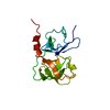
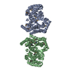
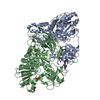
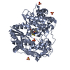
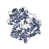
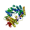
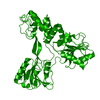
Entry Database : PDB / ID : 2wrmTitle Identification of Novel Allosteric Inhibitors of Hepatitis C Virus NS5B Polymerase Thumb Domain (Site II) by Structure-Based Design RNA-DIRECTED RNA POLYMERASE Keywords / / / Function / homology Function Domain/homology Component
/ / / / / / / / / / / / / / / / / / / / / / / / / / / / / / / / / / / / / / / / / / / / / / / / / / / / / / / / / / / / / / / / / / / / / / / / / / / / / / / / / / / / / / / / / / / / / / / / / / / / / / / / / / / / / Biological species Method / / / Resolution : 1.95 Å Authors Di Marco, S. Journal : To be Published Title : Identification of Novel Allosteric Inhibitors of Hepatitis C Virus Ns5B Polymerase Thumb Domain (Site II) by Structure-Based DesignAuthors : Di Francesco, M.E. / Di Marco, S. / Summa, V. History Deposition Sep 1, 2009 Deposition site / Processing site Revision 1.0 Sep 22, 2010 Provider / Type Revision 1.1 May 8, 2011 Group Revision 1.2 Jul 13, 2011 Group Revision 1.3 Dec 20, 2023 Group Data collection / Database references ... Data collection / Database references / Derived calculations / Other / Refinement description Category chem_comp_atom / chem_comp_bond ... chem_comp_atom / chem_comp_bond / database_2 / pdbx_database_status / pdbx_initial_refinement_model / struct_site Item _database_2.pdbx_DOI / _database_2.pdbx_database_accession ... _database_2.pdbx_DOI / _database_2.pdbx_database_accession / _pdbx_database_status.status_code_sf / _struct_site.pdbx_auth_asym_id / _struct_site.pdbx_auth_comp_id / _struct_site.pdbx_auth_seq_id
Show all Show less
 Movie
Movie Controller

 Yorodumi
 Open data
 Basic information
 Components
 RNA-dependent RNA polymerase
  Keywords
 Function and homology information
 HEPATITIS C VIRUS
 Authors
 Citation
 Structure visualization
 Downloads & links
 Other downloads
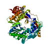


 PDBj
PDBj
 Assembly


 Mass: 326.347 Da / Num. of mol.: 3 / Source method: obtained synthetically / Formula: C18H18N2O4
 Mass: 18.015 Da / Num. of mol.: 629 / Source method: isolated from a natural source / Formula: H2O
 Sample preparation
 / Beamline: ID14-3 / Wavelength: 0.931
  Processing