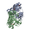
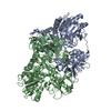
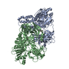
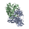
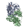
[English] 日本語
Yorodumi
- PDB-1cu1: CRYSTAL STRUCTURE OF AN ENZYME COMPLEX FROM HEPATITIS C VIRUS -

+
Open data


ID or keywords:

Loading...

-
Basic information

Entry
Database: PDB / ID: 1cu1
TitleCRYSTAL STRUCTURE OF AN ENZYME COMPLEX FROM HEPATITIS C VIRUS
ComponentsPROTEIN (PROTEASE/HELICASE NS3)
KeywordsHYDROLASE / hepatitis C Virus / bifunctional / protease-helicase
Function / homology
Function and homology information


hepacivirin / host cell mitochondrial membrane / host cell lipid droplet / symbiont-mediated suppression of host TRAF-mediated signal transduction / transformation of host cell by virus / symbiont-mediated perturbation of host cell cycle G1/S transition checkpoint / symbiont-mediated suppression of host JAK-STAT cascade via inhibition of STAT1 activity / symbiont-mediated suppression of host cytoplasmic pattern recognition receptor signaling pathway via inhibition of MAVS activity / SH3 domain binding / nucleoside-triphosphate phosphatase ...hepacivirin / host cell mitochondrial membrane / host cell lipid droplet / symbiont-mediated suppression of host TRAF-mediated signal transduction / transformation of host cell by virus / symbiont-mediated perturbation of host cell cycle G1/S transition checkpoint / symbiont-mediated suppression of host JAK-STAT cascade via inhibition of STAT1 activity / symbiont-mediated suppression of host cytoplasmic pattern recognition receptor signaling pathway via inhibition of MAVS activity / SH3 domain binding / nucleoside-triphosphate phosphatase / protein complex oligomerization / monoatomic ion channel activity / viral nucleocapsid / clathrin-dependent endocytosis of virus by host cell / Hydrolases; Acting on peptide bonds (peptidases); Cysteine endopeptidases / RNA helicase activity / host cell perinuclear region of cytoplasm / host cell endoplasmic reticulum membrane / symbiont-mediated suppression of host type I interferon-mediated signaling pathway / RNA helicase / ribonucleoprotein complex / induction by virus of host autophagy / RNA-directed RNA polymerase / virus-mediated perturbation of host defense response / viral RNA genome replication / cysteine-type endopeptidase activity / RNA-dependent RNA polymerase activity / serine-type endopeptidase activity / fusion of virus membrane with host endosome membrane / viral envelope / host cell nucleus / virion attachment to host cell / host cell plasma membrane / virion membrane / structural molecule activity / ATP hydrolysis activity / proteolysis / RNA binding / zinc ion binding / ATP binding / membrane
Similarity search - Function
RNA Helicase; Chain A, domain 3 / RNA Helicase Chain A , domain 3 / Thrombin, subunit H - #120 / : / Hepatitis C virus core protein, chain A superfamily / Hepatitus C virus, Non-structural 5a protein, C-terminal / Hepatitis C virus NS5A, 1B domain superfamily / Hepatitis C virus non-structural protein NS2, C-terminal domain / Hepatitis C virus non-structural protein NS2, N-terminal domain / Hepatitis C virus non-structural protein NS2 ...RNA Helicase; Chain A, domain 3 / RNA Helicase Chain A , domain 3 / Thrombin, subunit H - #120 / : / Hepatitis C virus core protein, chain A superfamily / Hepatitus C virus, Non-structural 5a protein, C-terminal / Hepatitis C virus NS5A, 1B domain superfamily / Hepatitis C virus non-structural protein NS2, C-terminal domain / Hepatitis C virus non-structural protein NS2, N-terminal domain / Hepatitis C virus non-structural protein NS2 / HCV NS5a protein C-terminal region / Hepatitis C virus, Non-structural protein NS4b / Hepatitis C virus, Core protein, N-terminal / Hepatitis C virus non-structural protein NS4b / Hepatitis C virus capsid protein / Hepatitis C virus, Non-structural protein NS2 / Hepatitis C virus, Non-structural 5a protein / Hepatitis C virus, Non-structural 5a protein, domain 1a / Hepatitis C virus non-structural 5a, 1B domain / NS5A domain 1a superfamily / Hepatitis C virus non-structural 5a protein membrane anchor / Hepatitis C virus non-structural 5a zinc finger domain / Hepatitis C virus non-structural 5a domain 1b / Hepacivirus nonstructural protein 2 (NS2) protease domain profile. / Hepatitis C virus, Non-structural protein NS4a / Hepatitis C virus non-structural protein NS4a / Hepatitis C virus, Core protein, C-terminal / Hepatitis C virus core protein / Hepatitis C virus, Non-structural protein E2/NS1 / Hepatitis C virus non-structural protein E2/NS1 / Hepatitis C virus, Envelope glycoprotein E1 / Hepatitis C virus envelope glycoprotein E1 / RNA dependent RNA polymerase, hepatitis C virus / Viral RNA dependent RNA polymerase / Hepatitis C virus, NS3 protease, Peptidase S29 / Hepatitis C virus NS3 protease / Hepacivirus/Pegivirus NS3 protease domain profile. / DEAD box, Flavivirus / Flavivirus DEAD domain / Trypsin-like serine proteases / Superfamilies 1 and 2 helicase C-terminal domain profile. / Superfamilies 1 and 2 helicase ATP-binding type-1 domain profile. / DEAD-like helicases superfamily / Helicase, C-terminal / Helicase superfamily 1/2, ATP-binding domain / Thrombin, subunit H / P-loop containing nucleotide triphosphate hydrolases / Reverse transcriptase/Diguanylate cyclase domain / RNA-directed RNA polymerase, catalytic domain / RdRp of positive ssRNA viruses catalytic domain profile. / Peptidase S1, PA clan, chymotrypsin-like fold / Peptidase S1, PA clan / DNA/RNA polymerase superfamily / Beta Barrel / P-loop containing nucleoside triphosphate hydrolase / Rossmann fold / Orthogonal Bundle / 3-Layer(aba) Sandwich / Mainly Beta / Mainly Alpha / Alpha Beta
Similarity search - Domain/homology
PHOSPHATE ION / Genome polyprotein
Similarity search - Component
Biological speciesHepatitis C virus
MethodX-RAY DIFFRACTION / SYNCHROTRON / Resolution: 2.5 Å
AuthorsYao, N. / Weber, P.C.
CitationJournal: Structure Fold.Des. / Year: 1999
Title: Molecular views of viral polyprotein processing revealed by the crystal structure of the hepatitis C virus bifunctional protease-helicase.
Authors: Yao, N. / Reichert, P. / Taremi, S.S. / Prosise, W.W. / Weber, P.C.
History
DepositionAug 20, 1999Deposition site: RCSB / Processing site: RCSB
Revision 1.0Aug 23, 2000Provider: repository / Type: Initial release
Revision 1.1Apr 27, 2008Group: Version format compliance
Revision 1.2Jul 13, 2011Group: Version format compliance
Revision 1.3Aug 23, 2017Group: Source and taxonomy / Category: entity_src_gen
Revision 1.4Feb 7, 2024Group: Data collection / Database references / Derived calculations
Category: chem_comp_atom / chem_comp_bond ...chem_comp_atom / chem_comp_bond / database_2 / struct_conn / struct_site
Item: _database_2.pdbx_DOI / _database_2.pdbx_database_accession ..._database_2.pdbx_DOI / _database_2.pdbx_database_accession / _struct_conn.pdbx_dist_value / _struct_conn.ptnr1_auth_asym_id / _struct_conn.ptnr1_auth_comp_id / _struct_conn.ptnr1_auth_seq_id / _struct_conn.ptnr1_label_asym_id / _struct_conn.ptnr1_label_atom_id / _struct_conn.ptnr1_label_comp_id / _struct_conn.ptnr1_label_seq_id / _struct_conn.ptnr2_auth_asym_id / _struct_conn.ptnr2_auth_comp_id / _struct_conn.ptnr2_auth_seq_id / _struct_conn.ptnr2_label_asym_id / _struct_conn.ptnr2_label_atom_id / _struct_conn.ptnr2_label_comp_id / _struct_site.pdbx_auth_asym_id / _struct_site.pdbx_auth_comp_id / _struct_site.pdbx_auth_seq_id

-
Structure visualization

Structure viewerMolecule:
MolmilJmol/JSmol

Downloads & links

-
Assembly

Deposited unit
A: PROTEIN (PROTEASE/HELICASE NS3)
B: PROTEIN (PROTEASE/HELICASE NS3)
hetero molecules


Theoretical massNumber of molelcules
Total (without water)137,4546
Polymers137,1342
Non-polymers3214
Water4,828268
1


  • Idetical with deposited unit
  • defined by author&software
TypeNameSymmetry operationNumber
identity operation1_555x,y,z1
Buried area3130 Å2
ΔGint-104 kcal/mol
Surface area51330 Å2
MethodPISA
Unit cell
Length a, b, c (Å)91.36, 110.51, 141.20
Angle α, β, γ (deg.)90, 90, 90
Int Tables number19
Space group name H-MP212121

-
Components

#1: Protein PROTEIN (PROTEASE/HELICASE NS3)


Mass: 68566.828 Da / Num. of mol.: 2
Source method: isolated from a genetically manipulated source
Source: (gene. exp.) Hepatitis C virus / Genus: Hepacivirus / Production host: Escherichia coli (E. coli)
References: UniProt: P26663, Hydrolases; Acting on peptide bonds (peptidases); Serine endopeptidases
#2: Chemical ChemComp-ZN / ZINC ION


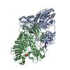
Mass: 65.409 Da / Num. of mol.: 2 / Source method: obtained synthetically / Formula: Zn
#3: Chemical ChemComp-PO4 / PHOSPHATE ION / Phosphate


Mass: 94.971 Da / Num. of mol.: 2 / Source method: obtained synthetically / Formula: PO4
#4: Water ChemComp-HOH / water / Water


Mass: 18.015 Da / Num. of mol.: 268 / Source method: isolated from a natural source / Formula: H2O

-
Experimental details

-
Experiment

ExperimentMethod: X-RAY DIFFRACTION / Number of used crystals: 1

-
Sample preparation

CrystalDensity Matthews: 2.6 Å3/Da / Density % sol: 52.64 %
Crystal grow
*PLUS
pH: 6.4 / Method: batch method
Components of the solutions
*PLUS
IDConc.Common nameCrystal-IDSol-IDChemical formula
10.07 mMprotein11
20.08 Msodium phosphate11
316 %(w/v)PEG600011
432 mMdi-potassium hydrogen phosphate11
58 %(w/v)MPD11
65 mMHEPES11
70.2 M11NaCl
82 %(v/v)glycerol11
92 mMbeta-mercaptoethanol11

-
Data collection

Diffraction sourceSource: SYNCHROTRON / Site: APS / Type: APS
DetectorType: MARRESEARCH / Detector: CCD
RadiationProtocol: SINGLE WAVELENGTH / Monochromatic (M) / Laue (L): M / Scattering type: x-ray
Radiation wavelengthRelative weight: 1
ReflectionResolution: 2.5→40 Å / Num. all: 50404 / Num. obs: 50441 / % possible obs: 100 % / Observed criterion σ(F): 0 / Observed criterion σ(I): 0 / Redundancy: 5.6 % / Rmerge(I) obs: 0.082 / Net I/σ(I): 21
Reflection shellResolution: 2.5→2.59 Å / Redundancy: 5.6 % / Rmerge(I) obs: 0.34 / % possible all: 100
Reflection
*PLUS
Num. measured all: 601477

-
Processing

Software
NameVersionClassification
X-PLORmodel building
X-PLOR98.1refinement
DENZOdata reduction
SCALEPACKdata scaling
X-PLORphasing
RefinementResolution: 2.5→40 Å / σ(F): 2
RfactorNum. reflection
Rfree0.26 1003
Rwork0.2 -
obs0.2 50404
all-50441
Refinement stepCycle: LAST / Resolution: 2.5→40 Å
ProteinNucleic acidLigandSolventTotal
Num. atoms9614 0 12 268 9894
Refine LS restraints
Refine-IDTypeDev ideal
X-RAY DIFFRACTIONx_bond_d0.013
X-RAY DIFFRACTIONx_bond_d_na
X-RAY DIFFRACTIONx_bond_d_prot
X-RAY DIFFRACTIONx_angle_d
X-RAY DIFFRACTIONx_angle_d_na
X-RAY DIFFRACTIONx_angle_d_prot
X-RAY DIFFRACTIONx_angle_deg1.67
X-RAY DIFFRACTIONx_angle_deg_na
X-RAY DIFFRACTIONx_angle_deg_prot
X-RAY DIFFRACTIONx_dihedral_angle_d
X-RAY DIFFRACTIONx_dihedral_angle_d_na
X-RAY DIFFRACTIONx_dihedral_angle_d_prot
X-RAY DIFFRACTIONx_improper_angle_d
X-RAY DIFFRACTIONx_improper_angle_d_na
X-RAY DIFFRACTIONx_improper_angle_d_prot
X-RAY DIFFRACTIONx_mcbond_it
X-RAY DIFFRACTIONx_mcangle_it
X-RAY DIFFRACTIONx_scbond_it
X-RAY DIFFRACTIONx_scangle_it
Software
*PLUS
Name: X-PLOR / Version: 98.1 / Classification: refinement
Refinement
*PLUS
Highest resolution: 2.5 Å / Lowest resolution: 40 Å / σ(F): 2 / Rfactor obs: 0.2 / Rfactor Rfree: 0.26 / Rfactor Rwork: 0.2
Solvent computation
*PLUS
Displacement parameters
*PLUS

+
About Yorodumi

-
News

-
Feb 9, 2022. New format data for meta-information of EMDB entries

New format data for meta-information of EMDB entries

  • Version 3 of the EMDB header file is now the official format.
  • The previous official version 1.9 will be removed from the archive.

Related info.:EMDB header

External links:wwPDB to switch to version 3 of the EMDB data model

-
Aug 12, 2020. Covid-19 info

Covid-19 info

URL: https://pdbj.org/emnavi/covid19.php

New page: Covid-19 featured information page in EM Navigator.

Related info.:Covid-19 info / Mar 5, 2020. Novel coronavirus structure data

+
Mar 5, 2020. Novel coronavirus structure data

Novel coronavirus structure data

Related info.:Yorodumi Speices / Aug 12, 2020. Covid-19 info

External links:COVID-19 featured content - PDBj / Molecule of the Month (242):Coronavirus Proteases

+
Jan 31, 2019. EMDB accession codes are about to change! (news from PDBe EMDB page)

EMDB accession codes are about to change! (news from PDBe EMDB page)

  • The allocation of 4 digits for EMDB accession codes will soon come to an end. Whilst these codes will remain in use, new EMDB accession codes will include an additional digit and will expand incrementally as the available range of codes is exhausted. The current 4-digit format prefixed with “EMD-” (i.e. EMD-XXXX) will advance to a 5-digit format (i.e. EMD-XXXXX), and so on. It is currently estimated that the 4-digit codes will be depleted around Spring 2019, at which point the 5-digit format will come into force.
  • The EM Navigator/Yorodumi systems omit the EMD- prefix.

Related info.:Q: What is EMD? / ID/Accession-code notation in Yorodumi/EM Navigator

External links:EMDB Accession Codes are Changing Soon! / Contact to PDBj

+
Jul 12, 2017. Major update of PDB

Major update of PDB

  • wwPDB released updated PDB data conforming to the new PDBx/mmCIF dictionary.
  • This is a major update changing the version number from 4 to 5, and with Remediation, in which all the entries are updated.
  • In this update, many items about electron microscopy experimental information are reorganized (e.g. em_software).
  • Now, EM Navigator and Yorodumi are based on the updated data.

External links:wwPDB Remediation / Enriched Model Files Conforming to OneDep Data Standards Now Available in the PDB FTP Archive

-
Yorodumi

Thousand views of thousand structures

  • Yorodumi is a browser for structure data from EMDB, PDB, SASBDB, etc.
  • This page is also the successor to EM Navigator detail page, and also detail information page/front-end page for Omokage search.
  • The word "yorodu" (or yorozu) is an old Japanese word meaning "ten thousand". "mi" (miru) is to see.

Related info.:EMDB / PDB / SASBDB / Comparison of 3 databanks / Yorodumi Search / Aug 31, 2016. New EM Navigator & Yorodumi / Yorodumi Papers / Jmol/JSmol / Function and homology information / Changes in new EM Navigator and Yorodumi

Read more