 Movie
Movie Controller

[English] 日本語
 Yorodumi
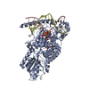
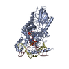
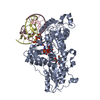
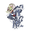
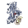
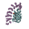
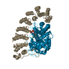
- PDB-2wq7: Structure of the 6-4 photolyase of D. melanogaster in complex wit... -
+ Open data

- Basic information
| Entry | Database: PDB / ID: 2wq7 | |||||||||
|---|---|---|---|---|---|---|---|---|---|---|
| Title | Structure of the 6-4 photolyase of D. melanogaster in complex with the non-natural N4-methyl T(6-4)C lesion | |||||||||
 Components Components |
| |||||||||
 Keywords Keywords | LYASE/DNA / LYASE-DNA COMPLEX /  DNA REPAIR / DNA LESION / LYASE DNA REPAIR / DNA LESION / LYASE | |||||||||
| Function / homology |  Function and homology informationdeoxyribodipyrimidine photo-lyase activity / entrainment of circadian clock by photoperiod / FAD binding / circadian regulation of gene expression / DNA binding / nucleus / cytoplasm Function and homology informationdeoxyribodipyrimidine photo-lyase activity / entrainment of circadian clock by photoperiod / FAD binding / circadian regulation of gene expression / DNA binding / nucleus / cytoplasmSimilarity search - Function | |||||||||
| Biological species |  DROSOPHILA MELANOGASTER (fruit fly) DROSOPHILA MELANOGASTER (fruit fly) | |||||||||
| Method | X-RAY DIFFRACTION / SYNCHROTRON / MOLECULAR REPLACEMENT / Resolution: 2 Å | |||||||||
 Authors Authors | Glas, A.F. / Kaya, E. / Schneider, S. / Maul, M.J. / Carell, T. | |||||||||
 Citation Citation | Journal: J.Am.Chem.Soc. / Year: 2010 Title: DNA (6-4) Photolyases Reduce Dewar Isomers for Isomerization Into (6-4) Lesions. Authors: Glas, A.F. / Kaya, E. / Schneider, S. / Heil, K. / Fazio, D. / Maul, M.J. / Carell, T. | |||||||||
| History |
|
- Structure visualization
| Structure viewer | Molecule: MolmilJmol/JSmol |
|---|
- Downloads & links
-Download
| PDBx/mmCIF format | 2wq7.cif.gz | 149.5 KB | Display | PDBx/mmCIF format |
|---|---|---|---|---|
| PDB format | pdb2wq7.ent.gz | 110.8 KB | Display | PDB format |
| PDBx/mmJSON format | 2wq7.json.gz | Tree view | PDBx/mmJSON format | |
| Others |  Other downloads Other downloads |
-Validation report
| Arichive directory | https://data.pdbj.org/pub/pdb/validation_reports/wq/2wq7ftp://data.pdbj.org/pub/pdb/validation_reports/wq/2wq7 | HTTPS FTP |
|---|
-Related structure data
| Related structure data |  2wq6C  3cvuS C: citing same article ( S: Starting model for refinement |
|---|---|
| Similar structure data |


-Links
 PDBj
PDBj


- Assembly
| Deposited unit | 
| ||||||||
|---|---|---|---|---|---|---|---|---|---|
| 1 |
| ||||||||
| Unit cell |
|
-Components
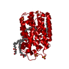
| #1: Protein | Mass: 62910.137 Da / Num. of mol.: 1 / Fragment: RESIDUES 1-520 Source method: isolated from a genetically manipulated source Source: (gene. exp.) DROSOPHILA MELANOGASTER (fruit fly) / Plasmid: PDEST007 DERIVATIVE / Production host:  ESCHERICHIA COLI (E. coli) / Strain (production host): ROSETTA-GAMI PLYSS (DE3) ESCHERICHIA COLI (E. coli) / Strain (production host): ROSETTA-GAMI PLYSS (DE3)References: UniProt: Q8SXK5, deoxyribodipyrimidine photo-lyase |
|---|---|
| #2: DNA chain | Mass: 4651.054 Da / Num. of mol.: 1 / Source method: obtained synthetically |
| #3: DNA chain | Mass: 4545.948 Da / Num. of mol.: 1 / Source method: obtained synthetically |
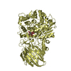
| #4: Chemical | ChemComp-FAD / Flavin adenine dinucleotide  Mass: 785.550 Da / Num. of mol.: 1 / Source method: obtained synthetically / Formula: C27H33N9O15P2 / Comment: FAD*YM Mass: 785.550 Da / Num. of mol.: 1 / Source method: obtained synthetically / Formula: C27H33N9O15P2 / Comment: FAD*YM |
| #5: Water | ChemComp-HOH / Water Mass: 18.015 Da / Num. of mol.: 403 / Source method: isolated from a natural source / Formula: H2O Mass: 18.015 Da / Num. of mol.: 403 / Source method: isolated from a natural source / Formula: H2O |
-Experimental details
-Experiment
| Experiment | Method: X-RAY DIFFRACTION / Number of used crystals: 1 |
|---|
- Sample preparation
| Crystal | Density Matthews: 2.95 Å3/Da / Density % sol: 58 % / Description: NONE |
|---|---|
| Crystal grow | Details: 100MM HEPES PH 7, 13-17% PEG1500 18DGC |
-Data collection
| Diffraction | Mean temperature: 100 K |
|---|---|
| Diffraction source | Source: SYNCHROTRON / Site: SLS  / Beamline: X10SA / Wavelength: 1 / Beamline: X10SA / Wavelength: 1 |
| Detector | Type: MARMOSAIC 225 mm CCD / Detector: CCD / Date: Jul 27, 2009 |
| Radiation | Protocol: SINGLE WAVELENGTH / Monochromatic (M) / Laue (L): M / Scattering type: x-ray |
| Radiation wavelength | Wavelength: 1 Å / Relative weight: 1 |
| Reflection | Resolution: 2→40.5 Å / Num. obs: 48536 / % possible obs: 99.2 % / Observed criterion σ(I): 2 / Redundancy: 3.6 % / Biso Wilson estimate: 26.67 Å2 / Rmerge(I) obs: 0.06 / Net I/σ(I): 14.7 |
| Reflection shell | Resolution: 2→2.1 Å / Redundancy: 3.6 % / Rmerge(I) obs: 0.38 / Mean I/σ(I) obs: 3.7 / % possible all: 96.2 |
- Processing
| Software |
| |||||||||||||||||||||||||||||||||||||||||||||||||||||||||||||||||||||||||||||||||||||||||||||||||||||||||||||||||||||||||||||||||||||
|---|---|---|---|---|---|---|---|---|---|---|---|---|---|---|---|---|---|---|---|---|---|---|---|---|---|---|---|---|---|---|---|---|---|---|---|---|---|---|---|---|---|---|---|---|---|---|---|---|---|---|---|---|---|---|---|---|---|---|---|---|---|---|---|---|---|---|---|---|---|---|---|---|---|---|---|---|---|---|---|---|---|---|---|---|---|---|---|---|---|---|---|---|---|---|---|---|---|---|---|---|---|---|---|---|---|---|---|---|---|---|---|---|---|---|---|---|---|---|---|---|---|---|---|---|---|---|---|---|---|---|---|---|---|---|
| Refinement | Method to determine structure: MOLECULAR REPLACEMENT Starting model: PDB ENTRY 3CVU Resolution: 2→40.468 Å / SU ML: 0.43 / σ(F): 1.35 / Phase error: 19.97 / Stereochemistry target values: ML Details: RESIDUES DT 8 AND DC 9 HAVE FORMED A 6-4 PHOTOPRODUCT BY THE COVALENT BONDING OF ATOM C6 OF TDY TO C4 OF Z.
| |||||||||||||||||||||||||||||||||||||||||||||||||||||||||||||||||||||||||||||||||||||||||||||||||||||||||||||||||||||||||||||||||||||
| Solvent computation | Shrinkage radii: 0.9 Å / VDW probe radii: 1.11 Å / Solvent model: FLAT BULK SOLVENT MODEL / Bsol: 40 Å2 / ksol: 0.351 e/Å3 | |||||||||||||||||||||||||||||||||||||||||||||||||||||||||||||||||||||||||||||||||||||||||||||||||||||||||||||||||||||||||||||||||||||
| Displacement parameters | Biso mean: 28.4 Å2
| |||||||||||||||||||||||||||||||||||||||||||||||||||||||||||||||||||||||||||||||||||||||||||||||||||||||||||||||||||||||||||||||||||||
| Refinement step | Cycle: LAST / Resolution: 2→40.468 Å
| |||||||||||||||||||||||||||||||||||||||||||||||||||||||||||||||||||||||||||||||||||||||||||||||||||||||||||||||||||||||||||||||||||||
| Refine LS restraints |
| |||||||||||||||||||||||||||||||||||||||||||||||||||||||||||||||||||||||||||||||||||||||||||||||||||||||||||||||||||||||||||||||||||||
| LS refinement shell |
|
