 Movie
Movie Controller


+ Open data

- Basic information
| Entry | Database: PDB / ID: 2wmz | ||||||
|---|---|---|---|---|---|---|---|
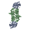
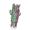
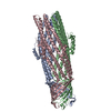
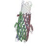
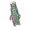
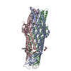
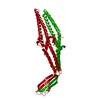
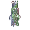
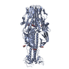
| Title | Structure of a mutated TolC | ||||||
 Components Components | OUTER MEMBRANE PROTEIN TOLC | ||||||
 Keywords Keywords |  MEMBRANE PROTEIN / TYPE-I SECRETION / CELL OUTER MEMBRANE / TRANSPORT / DRUG-EFFLUX MEMBRANE PROTEIN / TYPE-I SECRETION / CELL OUTER MEMBRANE / TRANSPORT / DRUG-EFFLUX | ||||||
| Function / homology |  Function and homology information Function and homology informationMacAB-TolC complex / enterobactin transport / bile acid transmembrane transporter activity / xenobiotic detoxification by transmembrane export across the cell outer membrane / efflux pump complex / periplasmic side of plasma membrane / xenobiotic detoxification by transmembrane export across the plasma membrane / bile acid and bile salt transport / porin activity / efflux transmembrane transporter activity ...MacAB-TolC complex / enterobactin transport / bile acid transmembrane transporter activity / xenobiotic detoxification by transmembrane export across the cell outer membrane / efflux pump complex / periplasmic side of plasma membrane / xenobiotic detoxification by transmembrane export across the plasma membrane / bile acid and bile salt transport / porin activity / efflux transmembrane transporter activity / monoatomic ion transmembrane transport / cell outer membrane / response to organic cyclic compound / response to toxic substance / monoatomic ion channel activity / outer membrane-bounded periplasmic space / response to xenobiotic stimulus / response to antibiotic / membrane / identical protein bindingSimilarity search - Function | ||||||
| Biological species |  ESCHERICHIA COLI (E. coli) ESCHERICHIA COLI (E. coli) | ||||||
| Method | X-RAY DIFFRACTION / SYNCHROTRON / MOLECULAR REPLACEMENT / Resolution: 2.9 Å | ||||||
 Authors Authors | Hinchliffe, P. / Pei, X.Y. / Symmons, M.F. / Koronakis, E. / Hughes, C. / Koronakis, V. | ||||||
 Citation Citation | Journal: Proc.Natl.Acad.Sci.USA / Year: 2011 Title: Structures of Sequential Open States in a Symmetrical Opening Transition of the Tolc Exit Duct. Authors: Pei, X.Y. / Hinchliffe, P. / Symmons, M.F. / Koronakis, E. / Benz, R. / Hughes, C. / Koronakis, V. | ||||||
| History |
| ||||||
| Remark 700 | SHEET DETERMINATION METHOD: DSSP THE SHEETS PRESENTED AS "AA" IN EACH CHAIN ON SHEET RECORDS BELOW ... SHEET DETERMINATION METHOD: DSSP THE SHEETS PRESENTED AS "AA" IN EACH CHAIN ON SHEET RECORDS BELOW IS ACTUALLY AN 13-STRANDED BARREL THIS IS REPRESENTED BY A 14-STRANDED SHEET IN WHICH THE FIRST AND LAST STRANDS ARE IDENTICAL. |
- Structure visualization
| Structure viewer | Molecule: MolmilJmol/JSmol |
|---|
- Downloads & links
-Download
| PDBx/mmCIF format | 2wmz.cif.gz | 252.1 KB | Display | PDBx/mmCIF format |
|---|---|---|---|---|
| PDB format | pdb2wmz.ent.gz | 205.9 KB | Display | PDB format |
| PDBx/mmJSON format | 2wmz.json.gz | Tree view | PDBx/mmJSON format | |
| Others |  Other downloads Other downloads |
-Validation report
| Arichive directory | https://data.pdbj.org/pub/pdb/validation_reports/wm/2wmzftp://data.pdbj.org/pub/pdb/validation_reports/wm/2wmz | HTTPS FTP |
|---|
-Related structure data
| Related structure data |  2xmnC 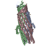 1ek9S S: Starting model for refinement C: citing same article ( |
|---|---|
| Similar structure data |


-Links
 PDBj
PDBj
- Assembly
| Deposited unit | 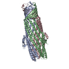
| ||||||||||||||||||||||||||||||||||||||||||
|---|---|---|---|---|---|---|---|---|---|---|---|---|---|---|---|---|---|---|---|---|---|---|---|---|---|---|---|---|---|---|---|---|---|---|---|---|---|---|---|---|---|---|---|
| 1 |
| ||||||||||||||||||||||||||||||||||||||||||
| Unit cell |
| ||||||||||||||||||||||||||||||||||||||||||
| Noncrystallographic symmetry (NCS) | NCS domain:
NCS oper:
|
-Components
| #1: Protein | Mass: 46933.723 Da / Num. of mol.: 3 Fragment: MATURE PROTEIN WITH 43 C-TERMINAL RESIDUES REMOVED, RESIDUES 23-450 Mutation: YES Source method: isolated from a genetically manipulated source Source: (gene. exp.) ESCHERICHIA COLI (E. coli) / Strain: K-12 / Plasmid: PT7TOLC / Production host: ESCHERICHIA COLI (E. coli) / Strain (production host): BL923(DE3) / Variant (production host): TOLC- / References: UniProt: P02930#2: Chemical | Chloride  Mass: 35.453 Da / Num. of mol.: 3 / Source method: obtained synthetically / Formula: Cl Mass: 35.453 Da / Num. of mol.: 3 / Source method: obtained synthetically / Formula: Cl#3: Chemical | ChemComp-PGE / Polyethylene glycol  Mass: 150.173 Da / Num. of mol.: 5 / Source method: obtained synthetically / Formula: C6H14O4 Mass: 150.173 Da / Num. of mol.: 5 / Source method: obtained synthetically / Formula: C6H14O4#4: Chemical | ChemComp-NA / |   Mass: 22.990 Da / Num. of mol.: 1 / Source method: obtained synthetically / Formula: Na Mass: 22.990 Da / Num. of mol.: 1 / Source method: obtained synthetically / Formula: Na#5: Water | ChemComp-HOH / | Water Mass: 18.015 Da / Num. of mol.: 24 / Source method: isolated from a natural source / Formula: H2O Mass: 18.015 Da / Num. of mol.: 24 / Source method: isolated from a natural source / Formula: H2OCompound details | ENGINEERED RESIDUE IN CHAIN A, ARG 389 TO SER ENGINEERED RESIDUE IN CHAIN B, ARG 389 TO SER ...ENGINEERED | Nonpolymer details | TRIETHYLEN | Sequence details | V TO L AT GENE POSITION 191 IN ISOLATE USED FOR STRUCTURE DETERMINATION - CORRESPONDS TO L169 IN ...V TO L AT GENE POSITION 191 IN ISOLATE USED FOR STRUCTURE DETERMINAT | |
|---|
-Experimental details
-Experiment
| Experiment | Method: X-RAY DIFFRACTION / Number of used crystals: 1 |
|---|
- Sample preparation
| Crystal | Density Matthews: 4.059 Å3/Da / Density % sol: 69 % / Description: NONE |
|---|---|
| Crystal grow | pH: 7 Details: 50 MM TRIS PH 8.2, 50 MM NACL, 14% PEG2000, 0.075% N-DODECYL-BETA-D-MALTOSIDE, 0.3% N-OCTYL BETA-D-GLUCOPYRANOSIDE |
-Data collection
| Diffraction | Mean temperature: 100 K |
|---|---|
| Diffraction source | Source: SYNCHROTRON / Site: Diamond  / Beamline: I02 / Wavelength: 0.9795 / Beamline: I02 / Wavelength: 0.9795 |
| Detector | Type: ADSC CCD / Detector: CCD / Date: Mar 3, 2009 / Details: KIRKPATRICK BAEZ BIMORPH MIRROR PAIR |
| Radiation | Monochromator: SI (111) DOUBLE CRYSTAL / Protocol: SINGLE WAVELENGTH / Monochromatic (M) / Laue (L): M / Scattering type: x-ray |
| Radiation wavelength | Wavelength: 0.9795 Å / Relative weight: 1 |
| Reflection | Resolution: 2.9→29.93 Å / Num. obs: 51195 / % possible obs: 98.9 % / Observed criterion σ(I): 0 / Redundancy: 5.8 % / Biso Wilson estimate: 61.8 Å2 / Rmerge(I) obs: 0.12 / Net I/σ(I): 11.1 |
| Reflection shell | Resolution: 2.9→3.1 Å / Rmerge(I) obs: 0.49 / Mean I/σ(I) obs: 3.8 / % possible all: 98 |
- Processing
| Software |
| ||||||||||||||||||||||||||||||||||||||||||||||||||||||||||||
|---|---|---|---|---|---|---|---|---|---|---|---|---|---|---|---|---|---|---|---|---|---|---|---|---|---|---|---|---|---|---|---|---|---|---|---|---|---|---|---|---|---|---|---|---|---|---|---|---|---|---|---|---|---|---|---|---|---|---|---|---|---|
| Refinement | Method to determine structure: MOLECULAR REPLACEMENT Starting model: PDB ENTRY 1EK9 CHAIN A Resolution: 2.9→29.93 Å / Rfactor Rfree error: 0.006 / Data cutoff high absF: 2898038.02 / Data cutoff low absF: 0 / Cross valid method: THROUGHOUT / σ(F): 0 / Stereochemistry target values: MAXIMUM LIKELIHOOD Details: GEOMETRIC REFINEMENT WAS CARRIED OUT USING CNS VERSION 1.1. B-FACTOR REFINEMENT, BULK SOLVENT MODELLING, AND FINAL R-FACTORS ARE FROM PHENIX. TRIETHYLENE GLYCOL PARAMETER FILE AND TOPOLOGY ...Details: GEOMETRIC REFINEMENT WAS CARRIED OUT USING CNS VERSION 1.1. B-FACTOR REFINEMENT, BULK SOLVENT MODELLING, AND FINAL R-FACTORS ARE FROM PHENIX. TRIETHYLENE GLYCOL PARAMETER FILE AND TOPOLOGY FILE WERE FROM HIC-UP DATABASE.
| ||||||||||||||||||||||||||||||||||||||||||||||||||||||||||||
| Solvent computation | Solvent model: PHENIX FLAT BULK SOLVENT MODEL / Bsol: 24.73 Å2 / ksol: 0.28 e/Å3 | ||||||||||||||||||||||||||||||||||||||||||||||||||||||||||||
| Displacement parameters | Biso mean: 59.6 Å2
| ||||||||||||||||||||||||||||||||||||||||||||||||||||||||||||
| Refine analyze |
| ||||||||||||||||||||||||||||||||||||||||||||||||||||||||||||
| Refinement step | Cycle: LAST / Resolution: 2.9→29.93 Å
| ||||||||||||||||||||||||||||||||||||||||||||||||||||||||||||
| Refine LS restraints |
| ||||||||||||||||||||||||||||||||||||||||||||||||||||||||||||
| Refine LS restraints NCS |
| ||||||||||||||||||||||||||||||||||||||||||||||||||||||||||||
| LS refinement shell | Resolution: 2.9→3.08 Å / Rfactor Rfree error: 0.024 / Total num. of bins used: 6
| ||||||||||||||||||||||||||||||||||||||||||||||||||||||||||||
| Xplor file |
|
