 Movie
Movie Controller

[English] 日本語
 Yorodumi
- PDB-2whi: Crystal structure of Mycobacterium Tuberculosis Glutamine Synthet... -
+ Open data

- Basic information
| Entry | Database: PDB / ID: 2whi | ||||||
|---|---|---|---|---|---|---|---|
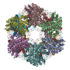
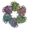
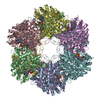
| Title | Crystal structure of Mycobacterium Tuberculosis Glutamine Synthetase in complex with a purine analogue inhibitor and L-methionine-S- sulfoximine phosphate. | ||||||
 Components Components | GLUTAMINE SYNTHETASE 1 | ||||||
 Keywords Keywords | LIGASE / NUCLEOTIDE-BINDING / TRANSITION STATE MIMIC / TAUT STATE / ATP-BINDING / PURINE ANALOGUE / GLNA1 / MT2278 / RV2220 / CYTOPLASM / SYNTHETASE | ||||||
| Function / homology |  Function and homology information Function and homology informationnitrogen utilization / positive regulation of plasminogen activation / glutamine synthetase / glutamine biosynthetic process / glutamine synthetase activity / zymogen binding / cell wall / fibronectin binding / peptidoglycan-based cell wall / protein homooligomerization ...nitrogen utilization / positive regulation of plasminogen activation / glutamine synthetase / glutamine biosynthetic process / glutamine synthetase activity / zymogen binding / cell wall / fibronectin binding / peptidoglycan-based cell wall / protein homooligomerization / magnesium ion binding / extracellular region / ATP binding / membrane / metal ion binding / plasma membrane / cytosol / cytoplasmSimilarity search - Function | ||||||
| Biological species |   MYCOBACTERIUM TUBERCULOSIS (bacteria) MYCOBACTERIUM TUBERCULOSIS (bacteria) | ||||||
| Method | X-RAY DIFFRACTION / SYNCHROTRON / MOLECULAR REPLACEMENT / Resolution: 2.2 Å | ||||||
 Authors Authors | Nilsson, M.T. / Krajewski, W.W. / Jones, T.A. / Mowbray, S.L. | ||||||
 Citation Citation | Journal: J.Mol.Biol. / Year: 2009 Title: Structural Basis for the Inhibition of Mycobacterium Tuberculosis Glutamine Synthetase by Novel ATP-Competitive Inhibitors. Authors: Nilsson, M.T. / Krajewski, W.W. / Yellagunda, S. / Prabhumurthy, S. / Chamarahally, G.N. / Siddamadappa, C. / Srinivasa, B.R. / Yahiaoui, S. / Larhed, M. / Karlen, A. / Jones, T.A. / Mowbray, S.L. #1: Journal: Proc.Natl.Acad.Sci.USA / Year: 2005Title: Structure of Mycobacterium Tuberculosis Glutamine Synthetase in Complex with a Transition-State Mimic Provides Functional Insights. Authors: Krajewski, W.W. / Jones, T.A. / Mowbray, S.L. | ||||||
| History |
| ||||||
| Remark 650 | HELIX DETERMINATION METHOD: AUTHOR PROVIDED. | ||||||
| Remark 700 | SHEET THE SHEET STRUCTURE OF THIS MOLECULE IS BIFURCATED. IN ORDER TO REPRESENT THIS FEATURE IN ... SHEET THE SHEET STRUCTURE OF THIS MOLECULE IS BIFURCATED. IN ORDER TO REPRESENT THIS FEATURE IN THE SHEET RECORDS BELOW, TWO SHEETS ARE DEFINED. |
- Structure visualization
| Structure viewer | Molecule: MolmilJmol/JSmol |
|---|
- Downloads & links
-Download
| PDBx/mmCIF format | 2whi.cif.gz | 597 KB | Display | PDBx/mmCIF format |
|---|---|---|---|---|
| PDB format | pdb2whi.ent.gz | 491.5 KB | Display | PDB format |
| PDBx/mmJSON format | 2whi.json.gz | Tree view | PDBx/mmJSON format | |
| Others |  Other downloads Other downloads |
-Validation report
| Arichive directory | https://data.pdbj.org/pub/pdb/validation_reports/wh/2whiftp://data.pdbj.org/pub/pdb/validation_reports/wh/2whi | HTTPS FTP |
|---|
-Related structure data
| Related structure data |  2wgsC  2bvcS C: citing same article ( S: Starting model for refinement |
|---|---|
| Similar structure data |


-Links
 PDBj
PDBj
- Assembly
| Deposited unit | 
| |||||||||||||||||||||||||||||||||||||||||||||||||||||||||||||||||||||||||||||||||||||||||||||||||||
|---|---|---|---|---|---|---|---|---|---|---|---|---|---|---|---|---|---|---|---|---|---|---|---|---|---|---|---|---|---|---|---|---|---|---|---|---|---|---|---|---|---|---|---|---|---|---|---|---|---|---|---|---|---|---|---|---|---|---|---|---|---|---|---|---|---|---|---|---|---|---|---|---|---|---|---|---|---|---|---|---|---|---|---|---|---|---|---|---|---|---|---|---|---|---|---|---|---|---|---|---|
| 1 | 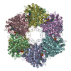
| |||||||||||||||||||||||||||||||||||||||||||||||||||||||||||||||||||||||||||||||||||||||||||||||||||
| 2 |
| |||||||||||||||||||||||||||||||||||||||||||||||||||||||||||||||||||||||||||||||||||||||||||||||||||
| Unit cell |
| |||||||||||||||||||||||||||||||||||||||||||||||||||||||||||||||||||||||||||||||||||||||||||||||||||
| Components on special symmetry positions |
| |||||||||||||||||||||||||||||||||||||||||||||||||||||||||||||||||||||||||||||||||||||||||||||||||||
| Noncrystallographic symmetry (NCS) | NCS domain:
NCS domain segments:
|
-Components
-Protein , 1 types, 6 molecules ABCDEF
| #1: Protein | / GLUTAMATE--AMMONIA LIGASE 1 Mass: 54584.730 Da / Num. of mol.: 6 / Fragment: RESIDUES 2-478 Source method: isolated from a genetically manipulated source Source: (gene. exp.) MYCOBACTERIUM TUBERCULOSIS (bacteria) / Strain: H37RV / Plasmid: PTRC99C / Production host: ESCHERICHIA COLI (E. coli) / Strain (production host): GJ4745References: UniProt: P0A590, UniProt: P9WN39*PLUS, glutamine synthetase |
|---|
-Non-polymers , 7 types, 1697 molecules 


| #2: Chemical | ChemComp-1AZ /  Mass: 424.281 Da / Num. of mol.: 6 / Source method: obtained synthetically / Formula: C18H19Cl2N5O3 Mass: 424.281 Da / Num. of mol.: 6 / Source method: obtained synthetically / Formula: C18H19Cl2N5O3#3: Chemical | ChemComp-MG /  Mass: 24.305 Da / Num. of mol.: 18 / Source method: obtained synthetically / Formula: Mg Mass: 24.305 Da / Num. of mol.: 18 / Source method: obtained synthetically / Formula: Mg#4: Chemical | ChemComp-P3S /  Mass: 260.205 Da / Num. of mol.: 6 / Source method: obtained synthetically / Formula: C5H13N2O6PS Mass: 260.205 Da / Num. of mol.: 6 / Source method: obtained synthetically / Formula: C5H13N2O6PS#5: Chemical | ChemComp-PO4 / Phosphate Mass: 94.971 Da / Num. of mol.: 6 / Source method: obtained synthetically / Formula: PO4 Mass: 94.971 Da / Num. of mol.: 6 / Source method: obtained synthetically / Formula: PO4#6: Chemical | ChemComp-CL / Chloride Mass: 35.453 Da / Num. of mol.: 6 / Source method: obtained synthetically / Formula: Cl Mass: 35.453 Da / Num. of mol.: 6 / Source method: obtained synthetically / Formula: Cl#7: Chemical | ChemComp-1PE / Polyethylene glycol Mass: 238.278 Da / Num. of mol.: 6 / Source method: obtained synthetically / Formula: C10H22O6 / Comment: precipitant*YM Mass: 238.278 Da / Num. of mol.: 6 / Source method: obtained synthetically / Formula: C10H22O6 / Comment: precipitant*YM#8: Water | ChemComp-HOH / | WaterMass: 18.015 Da / Num. of mol.: 1649 / Source method: isolated from a natural source / Formula: H2O |
|---|
-Details
| Sequence details | PROTEIN EXPRESSED IN FUSION WITH A N-TERMINAL 9 AMINO ACID PEPTIDE CONTAINING A SIX HISTIDINE ...PROTEIN EXPRESSED IN FUSION WITH A N-TERMINAL 9 AMINO ACID PEPTIDE CONTAINING |
|---|
-Experimental details
-Experiment
| Experiment | Method: X-RAY DIFFRACTION / Number of used crystals: 1 |
|---|
- Sample preparation
| Crystal | Density Matthews: 2.34 Å3/Da / Density % sol: 47.5 % Description: PROTEIN ONLY COORDINATES FROM THE ISOMORPHOUS CRYSTAL FORM FOUND IN PDB ENTRY 2BVC WERE USED AS THE STARTING POINT FOR REFINEMENT. |
|---|---|
| Crystal grow | Temperature: 294 K / Method: vapor diffusion, hanging drop / pH: 6.8 Details: EQUAL VOLUMES OF MOTHER LIQUOR (0.1 M MES, PH 6.8, 0.2 M MAGNESIUM CHLORIDE, 40% (V/V) PEG400) AND PROTEIN SOLUTION (30 G/L IN 0.1 M TRIS-HCL, PH 7.5, 0.125 M SODIUM CHLORIDE, 0.004 M ...Details: EQUAL VOLUMES OF MOTHER LIQUOR (0.1 M MES, PH 6.8, 0.2 M MAGNESIUM CHLORIDE, 40% (V/V) PEG400) AND PROTEIN SOLUTION (30 G/L IN 0.1 M TRIS-HCL, PH 7.5, 0.125 M SODIUM CHLORIDE, 0.004 M MAGNESIUM CHLORIDE, 4%(V/V) DMSO, 0.001 M INHIBITOR, 0.004 M MSOP), HANGING-DROP, VAPOR DIFFUSION, TEMPERATURE 294K. |
-Data collection
| Diffraction | Mean temperature: 100 K |
|---|---|
| Diffraction source | Source: SYNCHROTRON / Site: ESRF  / Beamline: ID14-1 / Wavelength: 0.934 / Beamline: ID14-1 / Wavelength: 0.934 |
| Detector | Type: ADSC CCD / Detector: CCD / Date: Feb 1, 2008 |
| Radiation | Protocol: SINGLE WAVELENGTH / Monochromatic (M) / Laue (L): M / Scattering type: x-ray |
| Radiation wavelength | Wavelength: 0.934 Å / Relative weight: 1 |
| Reflection | Resolution: 2.2→39.8 Å / Num. obs: 151747 / % possible obs: 97.2 % / Observed criterion σ(I): 0 / Redundancy: 4 % / Biso Wilson estimate: 31.2 Å2 / Rmerge(I) obs: 0.11 / Net I/σ(I): 13.4 |
| Reflection shell | Resolution: 2.2→2.3 Å / Redundancy: 2.8 % / Rmerge(I) obs: 0.34 / Mean I/σ(I) obs: 3.4 / % possible all: 86.5 |
- Processing
| Software |
| ||||||||||||||||||||||||||||||||||||||||||||||||||||||||||||||||||||||||||||||||||||||||||||||||||||||||||||||||||||||||||||||||||||||||||||||||||||||||||||||||||||||||||||||||||||||
|---|---|---|---|---|---|---|---|---|---|---|---|---|---|---|---|---|---|---|---|---|---|---|---|---|---|---|---|---|---|---|---|---|---|---|---|---|---|---|---|---|---|---|---|---|---|---|---|---|---|---|---|---|---|---|---|---|---|---|---|---|---|---|---|---|---|---|---|---|---|---|---|---|---|---|---|---|---|---|---|---|---|---|---|---|---|---|---|---|---|---|---|---|---|---|---|---|---|---|---|---|---|---|---|---|---|---|---|---|---|---|---|---|---|---|---|---|---|---|---|---|---|---|---|---|---|---|---|---|---|---|---|---|---|---|---|---|---|---|---|---|---|---|---|---|---|---|---|---|---|---|---|---|---|---|---|---|---|---|---|---|---|---|---|---|---|---|---|---|---|---|---|---|---|---|---|---|---|---|---|---|---|---|---|
| Refinement | Method to determine structure: MOLECULAR REPLACEMENT Starting model: PDB ENTRY 2BVC Resolution: 2.2→39.81 Å / Cor.coef. Fo:Fc: 0.932 / Cor.coef. Fo:Fc free: 0.922 / SU B: 7.307 / SU ML: 0.175 / Cross valid method: THROUGHOUT / ESU R: 0.346 / ESU R Free: 0.215 / Stereochemistry target values: MAXIMUM LIKELIHOOD Details: HYDROGENS HAVE BEEN ADDED IN THE RIDING POSITIONS. RESIDUES 403-416 ARE EXCLUDED FROM THE NCS DUE TO DIFFERENCES AS A RESULT OF CRYSTAL CONTACTS. E. COLI STRAIN GJ4745 IS ADENYLYLTRANSFERASE ...Details: HYDROGENS HAVE BEEN ADDED IN THE RIDING POSITIONS. RESIDUES 403-416 ARE EXCLUDED FROM THE NCS DUE TO DIFFERENCES AS A RESULT OF CRYSTAL CONTACTS. E. COLI STRAIN GJ4745 IS ADENYLYLTRANSFERASE DEFICIENT ( GLNE-) LONG CONTINUOUS DENSITY WAS MODELED AS PEG BASED ON THE HIGH PEG400 CONTENT IN THE CRYSTALLIZATION CONDITIONS.
| ||||||||||||||||||||||||||||||||||||||||||||||||||||||||||||||||||||||||||||||||||||||||||||||||||||||||||||||||||||||||||||||||||||||||||||||||||||||||||||||||||||||||||||||||||||||
| Solvent computation | Ion probe radii: 0.8 Å / Shrinkage radii: 0.8 Å / VDW probe radii: 1.2 Å / Solvent model: MASK | ||||||||||||||||||||||||||||||||||||||||||||||||||||||||||||||||||||||||||||||||||||||||||||||||||||||||||||||||||||||||||||||||||||||||||||||||||||||||||||||||||||||||||||||||||||||
| Displacement parameters | Biso mean: 27.424 Å2
| ||||||||||||||||||||||||||||||||||||||||||||||||||||||||||||||||||||||||||||||||||||||||||||||||||||||||||||||||||||||||||||||||||||||||||||||||||||||||||||||||||||||||||||||||||||||
| Refinement step | Cycle: LAST / Resolution: 2.2→39.81 Å
| ||||||||||||||||||||||||||||||||||||||||||||||||||||||||||||||||||||||||||||||||||||||||||||||||||||||||||||||||||||||||||||||||||||||||||||||||||||||||||||||||||||||||||||||||||||||
| Refine LS restraints |
|