 Movie
Movie Controller

[English] 日本語
 Yorodumi
- PDB-1hto: CRYSTALLOGRAPHIC STRUCTURE OF A RELAXED GLUTAMINE SYNTHETASE FROM... -
+ Open data

- Basic information
| Entry | Database: PDB / ID: 1hto | ||||||
|---|---|---|---|---|---|---|---|
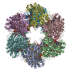
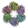
| Title | CRYSTALLOGRAPHIC STRUCTURE OF A RELAXED GLUTAMINE SYNTHETASE FROM MYCOBACTERIUM TUBERCULOSIS | ||||||
 Components Components | GLUTAMINE SYNTHETASE | ||||||
 Keywords Keywords | LIGASE / glutamine synthetase / Mycobacterium tuberculosis / Citrate / Structural Genomics / PSI / Protein Structure Initiative / TB Structural Genomics Consortium / TBSGC | ||||||
| Function / homology |  Function and homology information Function and homology informationnitrogen utilization / positive regulation of plasminogen activation / glutamine synthetase / glutamine biosynthetic process / glutamine synthetase activity / zymogen binding / fibronectin binding / peptidoglycan-based cell wall / protein homooligomerization / magnesium ion binding ...nitrogen utilization / positive regulation of plasminogen activation / glutamine synthetase / glutamine biosynthetic process / glutamine synthetase activity / zymogen binding / fibronectin binding / peptidoglycan-based cell wall / protein homooligomerization / magnesium ion binding / extracellular region / ATP binding / membrane / metal ion binding / plasma membrane / cytosol / cytoplasmSimilarity search - Function | ||||||
| Biological species |   Mycobacterium tuberculosis (bacteria) Mycobacterium tuberculosis (bacteria) | ||||||
| Method | X-RAY DIFFRACTION / SYNCHROTRON / MOLECULAR REPLACEMENT / Resolution: 2.4 Å | ||||||
 Authors Authors | Gill, H.S. / Eisenberg, D. / TB Structural Genomics Consortium (TBSGC) | ||||||
 Citation Citation | Journal: Biochemistry / Year: 2002 Title: Multicopy crystallographic refinement of a relaxed glutamine synthetase from Mycobacterium tuberculosis highlights flexible loops in the enzymatic mechanism and its regulation. Authors: Gill, H.S. / Pfluegl, G.M. / Eisenberg, D. | ||||||
| History |
|
- Structure visualization
| Structure viewer | Molecule: MolmilJmol/JSmol |
|---|
- Downloads & links
-Download
| PDBx/mmCIF format | 1hto.cif.gz | 2.3 MB | Display | PDBx/mmCIF format |
|---|---|---|---|---|
| PDB format | pdb1hto.ent.gz | 1.9 MB | Display | PDB format |
| PDBx/mmJSON format | 1hto.json.gz | Tree view | PDBx/mmJSON format | |
| Others |  Other downloads Other downloads |
-Validation report
| Arichive directory | https://data.pdbj.org/pub/pdb/validation_reports/ht/1htoftp://data.pdbj.org/pub/pdb/validation_reports/ht/1hto | HTTPS FTP |
|---|
-Related structure data
| Related structure data |  1htqC  1f52S C: citing same article ( S: Starting model for refinement |
|---|---|
| Similar structure data | |
| Other databases |


-Links
 PDBj
PDBj


- Assembly
| Deposited unit | 
| ||||||||
|---|---|---|---|---|---|---|---|---|---|
| 1 | 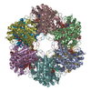
| ||||||||
| 2 | 
| ||||||||
| Unit cell |
| ||||||||
| Details | The biological complex is a dodecamer. |
-Components
| #1: Protein | / GS / GLUTAMATE--AMMONIA LIGASE 1 Mass: 53496.531 Da / Num. of mol.: 24 Source method: isolated from a genetically manipulated source Source: (gene. exp.) Mycobacterium tuberculosis (bacteria) / Plasmid: PTRCHISB / Production host: Escherichia coli (E. coli) / Strain (production host): YMC21EReferences: UniProt: Q10377, UniProt: P9WN39*PLUS, glutamine synthetase#2: Chemical | ChemComp-MN /   Mass: 54.938 Da / Num. of mol.: 24 / Source method: obtained synthetically / Formula: Mn Mass: 54.938 Da / Num. of mol.: 24 / Source method: obtained synthetically / Formula: Mn#3: Chemical | ChemComp-AMP / Adenosine monophosphate  Mass: 347.221 Da / Num. of mol.: 24 / Source method: obtained synthetically / Formula: C10H14N5O7P / Comment: AMP*YM Mass: 347.221 Da / Num. of mol.: 24 / Source method: obtained synthetically / Formula: C10H14N5O7P / Comment: AMP*YM#4: Chemical | ChemComp-CIT / Citric acid  Mass: 192.124 Da / Num. of mol.: 24 / Source method: obtained synthetically / Formula: C6H8O7 Mass: 192.124 Da / Num. of mol.: 24 / Source method: obtained synthetically / Formula: C6H8O7#5: Water | ChemComp-HOH / | Water Mass: 18.015 Da / Num. of mol.: 6312 / Source method: isolated from a natural source / Formula: H2O Mass: 18.015 Da / Num. of mol.: 6312 / Source method: isolated from a natural source / Formula: H2O |
|---|
-Experimental details
-Experiment
| Experiment | Method: X-RAY DIFFRACTION / Number of used crystals: 1 |
|---|
- Sample preparation
| Crystal | Density Matthews: 2.89 Å3/Da / Density % sol: 56.98 % |
|---|---|
| Crystal grow | Temperature: 298 K / Method: vapor diffusion, hanging drop / pH: 5.5 Details: PEG 4000, sodium citrate, sodium chloride, manganese chloride, imidazole buffer, pH 5.5, VAPOR DIFFUSION, HANGING DROP, temperature 298K |
-Data collection
| Diffraction | Mean temperature: 100 K |
|---|---|
| Diffraction source | Source: SYNCHROTRON / Site: NSLS  / Beamline: X12B / Wavelength: 0.978 Å / Beamline: X12B / Wavelength: 0.978 Å |
| Detector | Type: ADSC QUANTUM 4 / Detector: CCD / Date: Jul 1, 1997 |
| Radiation | Scattering type: x-ray |
| Radiation wavelength | Wavelength: 0.978 Å / Relative weight: 1 |
| Reflection | Resolution: 2.4→20 Å / Num. all: 566370 / Num. obs: 566370 / % possible obs: 99.6 % / Observed criterion σ(F): 0 / Observed criterion σ(I): 0 / Redundancy: 4.6 % / Biso Wilson estimate: 33.1 Å2 / Rmerge(I) obs: 0.075 / Net I/σ(I): 4.5 |
| Reflection shell | Resolution: 2.4→2.53 Å / Redundancy: 3.6 % / Rmerge(I) obs: 0.075 / Num. unique all: 80442 / % possible all: 98.8 |
- Processing
| Software |
| |||||||||||||||||||||||||
|---|---|---|---|---|---|---|---|---|---|---|---|---|---|---|---|---|---|---|---|---|---|---|---|---|---|---|
| Refinement | Method to determine structure: MOLECULAR REPLACEMENT Starting model: 1F52 Resolution: 2.4→20 Å / σ(F): 0 / σ(I): 0 / Stereochemistry target values: Engh & Huber Details: THE CRYSTAL PACKING WAS SOLVED USING MOLECULAR REPLACEMENT TECHNIQUES USING A DODECAMER MODEL FROM SALMONELLA TYPHIMURIUM GS AS A PROBE. THE MODEL WAS SUBSEQUENTLY REDUCED TO ONE SUBUNIT ...Details: THE CRYSTAL PACKING WAS SOLVED USING MOLECULAR REPLACEMENT TECHNIQUES USING A DODECAMER MODEL FROM SALMONELLA TYPHIMURIUM GS AS A PROBE. THE MODEL WAS SUBSEQUENTLY REDUCED TO ONE SUBUNIT WITH 24-FOLD NCS-SYMMETRICAL CONSTRAINTS IMPOSED. THE SUBUNIT WAS REFINED WITH STRICT 24-FOLD NCS-AVERAGING CONSTRAINTS AND THEN USED TO REGENERATE THE ENTIRE ASYMMETRIC UNIT. AS JUSTIFIED BY LOWER R-VALUES, THE FOLLOWING RESIDUES WERE COMPLETELY RELEASED AT THE END OF THE REFINEMENT IN ORDER ACCOMMODATE CRYSTAL CONTACTS: RESIDUES 385,98,602,11,7,40,3,292.
| |||||||||||||||||||||||||
| Refine analyze |
| |||||||||||||||||||||||||
| Refinement step | Cycle: LAST / Resolution: 2.4→20 Å
| |||||||||||||||||||||||||
| Refine LS restraints |
| |||||||||||||||||||||||||
| Xplor file | Serial no: 1 / Param file: PARHCSDX.PRO / Topol file: TOPHCSDX.PRO |
