Type: MARRESEARCH / Detector: CCD / Date: Jan 30, 2009
Radiation
Protocol: SINGLE WAVELENGTH / Monochromatic (M) / Laue (L): M / Scattering type: x-ray
Radiation wavelength
Wavelength: 0.88 Å / Relative weight: 1
Reflection
Resolution: 2.8→45.6 Å / Num. obs: 30800 / % possible obs: 100 % / Observed criterion σ(I): 2 / Redundancy: 3.7 % / Rmerge(I) obs: 0.09 / Net I/σ(I): 7.8
Reflection shell
Resolution: 2.8→2.95 Å / Redundancy: 3.7 % / Rmerge(I) obs: 0.53 / Mean I/σ(I) obs: 1.9 / % possible all: 100
-
Processing
Software
Name
Version
Classification
REFMAC
5.5.0085
refinement
MOSFLM
datareduction
SCALA
datascaling
PHASER
phasing
Refinement
Method to determine structure: MOLECULAR REPLACEMENT / Resolution: 2.8→45.6 Å / Cor.coef. Fo:Fc: 0.929 / Cor.coef. Fo:Fc free: 0.927 / SU B: 25.811 / SU ML: 0.255 / TLS residual ADP flag: LIKELY RESIDUAL / Cross valid method: THROUGHOUT / ESU R: 0.768 / ESU R Free: 0.326 / Stereochemistry target values: MAXIMUM LIKELIHOOD Details: HYDROGENS HAVE BEEN ADDED IN THE RIDING POSITIONS. UNK ATOMS RELATED TO A SECOND LIGAND WERE MODELED IN THE ACTIVE SITE.
Rfactor
Num. reflection
% reflection
Selection details
Rfree
0.248
1550
5 %
RANDOM
Rwork
0.224
-
-
-
obs
0.225
29219
99.9 %
-
Solvent computation
Ion probe radii: 0.8 Å / Shrinkage radii: 0.8 Å / VDW probe radii: 1.4 Å / Solvent model: MASK
 Movie
Movie Controller


 Open data
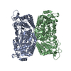
 Basic information
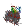
 Components
 Keywords
 OXIDOREDUCTASE /
  Function and homology information
 Authors
 Citation
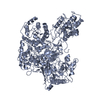
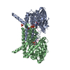
 Structure visualization
 Downloads & links
 Other downloads


 PDBj
PDBj
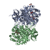
 Assembly


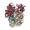
 Mass: 785.550 Da / Num. of mol.: 2 / Source method: obtained synthetically / Formula: C27H33N9O15P2 / Comment: FAD*YM
 Mass: 94.971 Da / Num. of mol.: 2 / Source method: obtained synthetically / Formula: PO4
 Num. of mol.: 14 / Source method: obtained synthetically
 Mass: 18.015 Da / Num. of mol.: 24 / Source method: isolated from a natural source / Formula: H2O
 Sample preparation
 / Beamline: X10SA / Wavelength: 0.88
  Processing