 Movie
Movie Controller


+ Open data

- Basic information
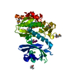
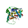
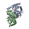
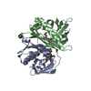
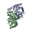
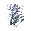
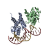
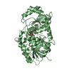
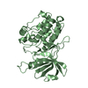
| Entry | Database: PDB / ID: 2wb8 | ||||||
|---|---|---|---|---|---|---|---|
| Title | Crystal structure of Haspin kinase | ||||||
 Components Components | SERINE/THREONINE-PROTEIN KINASE HASPIN | ||||||
 Keywords Keywords |  TRANSFERASE / HISTONE MODIFICATION / PHOSPHOPROTEIN / ATYPICAL KINASE / SERINE/THREONINE-PROTEIN KINASE / NUCLEOTIDE-BINDING / CHROMATIN REGULATOR / CELL CYCLE / HISTONE H3 / ATP-BINDING / GSG2 / KINASE / HASPIN / NUCLEUS / MITOSIS TRANSFERASE / HISTONE MODIFICATION / PHOSPHOPROTEIN / ATYPICAL KINASE / SERINE/THREONINE-PROTEIN KINASE / NUCLEOTIDE-BINDING / CHROMATIN REGULATOR / CELL CYCLE / HISTONE H3 / ATP-BINDING / GSG2 / KINASE / HASPIN / NUCLEUS / MITOSIS | ||||||
| Function / homology |  Function and homology information Function and homology informationhistone H3T3 kinase activity / protein localization to chromosome, centromeric region / mitotic sister chromatid cohesion / mitotic spindle assembly checkpoint signaling / spindle / chromosome / mitotic cell cycle / non-specific serine/threonine protein kinase / protein kinase activity / intracellular signal transduction ...histone H3T3 kinase activity / protein localization to chromosome, centromeric region / mitotic sister chromatid cohesion / mitotic spindle assembly checkpoint signaling / spindle / chromosome / mitotic cell cycle / non-specific serine/threonine protein kinase / protein kinase activity / intracellular signal transduction / protein phosphorylation / protein serine kinase activity / centrosome / nucleoplasm / ATP binding / nucleus / cytoplasmSimilarity search - Function | ||||||
| Biological species |  HOMO SAPIENS (human) HOMO SAPIENS (human) | ||||||
| Method | X-RAY DIFFRACTION / SYNCHROTRON / SAD / Resolution: 2.15 Å | ||||||
 Authors Authors | Villa, F. / Tortorci, M. / Forneris, F. / Mattevi, A. / Musacchio, A. | ||||||
 Citation Citation | Journal: Proc.Natl.Acad.Sci.USA / Year: 2009 Title: Crystal Structure of the Catalytic Domain of Haspin, an Atypical Kinase Implicated in Chromatin Organization. Authors: Villa, F. / Capasso, P. / Tortorici, M. / Forneris, F. / De Marco, A. / Mattevi, A. / Musacchio, A. | ||||||
| History |
|
- Structure visualization
| Structure viewer | Molecule: MolmilJmol/JSmol |
|---|
- Downloads & links
-Download
| PDBx/mmCIF format | 2wb8.cif.gz | 84.6 KB | Display | PDBx/mmCIF format |
|---|---|---|---|---|
| PDB format | pdb2wb8.ent.gz | 64 KB | Display | PDB format |
| PDBx/mmJSON format | 2wb8.json.gz | Tree view | PDBx/mmJSON format | |
| Others |  Other downloads Other downloads |
-Validation report
| Arichive directory | https://data.pdbj.org/pub/pdb/validation_reports/wb/2wb8ftp://data.pdbj.org/pub/pdb/validation_reports/wb/2wb8 | HTTPS FTP |
|---|
-Related structure data
| Related structure data | |
|---|---|
| Similar structure data |


-Links
 PDBj
PDBj- Assembly
| Deposited unit | 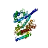
| ||||||||
|---|---|---|---|---|---|---|---|---|---|
| 1 |
| ||||||||
| Unit cell |
|
-Components
| #1: Protein | Mass: 39958.723 Da / Num. of mol.: 1 / Fragment: RESIDUES 452-798 Source method: isolated from a genetically manipulated source Source: (gene. exp.) HOMO SAPIENS (human) / Production host:  ESCHERICHIA COLI (E. coli) / Strain (production host): BL21 ESCHERICHIA COLI (E. coli) / Strain (production host): BL21References: UniProt: Q8TF76, non-specific serine/threonine protein kinase |
|---|---|
| #2: Water | ChemComp-HOH / Water Mass: 18.015 Da / Num. of mol.: 137 / Source method: isolated from a natural source / Formula: H2O Mass: 18.015 Da / Num. of mol.: 137 / Source method: isolated from a natural source / Formula: H2O |
-Experimental details
-Experiment
| Experiment | Method: X-RAY DIFFRACTION / Number of used crystals: 1 |
|---|
- Sample preparation
| Crystal | Density Matthews: 2.13 Å3/Da / Density % sol: 41.73 % / Description: NONE |
|---|
-Data collection
| Diffraction | Mean temperature: 101 K |
|---|---|
| Diffraction source | Source: SYNCHROTRON / Site: ESRF  / Beamline: ID14-1 / Wavelength: 0.9395 / Beamline: ID14-1 / Wavelength: 0.9395 |
| Detector | Type: ADSC CCD / Detector: CCD / Date: Jan 28, 2008 |
| Radiation | Protocol: SINGLE WAVELENGTH / Monochromatic (M) / Laue (L): M / Scattering type: x-ray |
| Radiation wavelength | Wavelength: 0.9395 Å / Relative weight: 1 |
| Reflection | Resolution: 2.15→50 Å / Num. obs: 18450 / % possible obs: 97.9 % / Observed criterion σ(I): 2 / Redundancy: 3.4 % / Rmerge(I) obs: 0.08 / Net I/σ(I): 15.8 |
| Reflection shell | Resolution: 2.15→2.2 Å / Redundancy: 3.3 % / Rmerge(I) obs: 0.48 / Mean I/σ(I) obs: 2.6 / % possible all: 98.1 |
- Processing
| Software |
| ||||||||||||||||||||||||||||||||||||||||||||||||||||||||||||||||||||||||||||||||||||||||||||||||||||||||||||||||||||||||||||||||||||||||||||||||||||||||||||||||||||||||||||||||||||||
|---|---|---|---|---|---|---|---|---|---|---|---|---|---|---|---|---|---|---|---|---|---|---|---|---|---|---|---|---|---|---|---|---|---|---|---|---|---|---|---|---|---|---|---|---|---|---|---|---|---|---|---|---|---|---|---|---|---|---|---|---|---|---|---|---|---|---|---|---|---|---|---|---|---|---|---|---|---|---|---|---|---|---|---|---|---|---|---|---|---|---|---|---|---|---|---|---|---|---|---|---|---|---|---|---|---|---|---|---|---|---|---|---|---|---|---|---|---|---|---|---|---|---|---|---|---|---|---|---|---|---|---|---|---|---|---|---|---|---|---|---|---|---|---|---|---|---|---|---|---|---|---|---|---|---|---|---|---|---|---|---|---|---|---|---|---|---|---|---|---|---|---|---|---|---|---|---|---|---|---|---|---|---|---|
| Refinement | Method to determine structure: SAD Starting model: NONE Resolution: 2.15→50 Å / Cor.coef. Fo:Fc: 0.941 / Cor.coef. Fo:Fc free: 0.91 / SU B: 16.369 / SU ML: 0.213 / TLS residual ADP flag: LIKELY RESIDUAL / Cross valid method: THROUGHOUT / ESU R: 0.331 / ESU R Free: 0.252 / Stereochemistry target values: MAXIMUM LIKELIHOOD / Details: HYDROGENS HAVE BEEN ADDED IN THE RIDING POSITIONS.
| ||||||||||||||||||||||||||||||||||||||||||||||||||||||||||||||||||||||||||||||||||||||||||||||||||||||||||||||||||||||||||||||||||||||||||||||||||||||||||||||||||||||||||||||||||||||
| Solvent computation | Ion probe radii: 0.8 Å / Shrinkage radii: 0.8 Å / VDW probe radii: 1.2 Å / Solvent model: MASK | ||||||||||||||||||||||||||||||||||||||||||||||||||||||||||||||||||||||||||||||||||||||||||||||||||||||||||||||||||||||||||||||||||||||||||||||||||||||||||||||||||||||||||||||||||||||
| Displacement parameters | Biso mean: 49.255 Å2
| ||||||||||||||||||||||||||||||||||||||||||||||||||||||||||||||||||||||||||||||||||||||||||||||||||||||||||||||||||||||||||||||||||||||||||||||||||||||||||||||||||||||||||||||||||||||
| Refinement step | Cycle: LAST / Resolution: 2.15→50 Å
| ||||||||||||||||||||||||||||||||||||||||||||||||||||||||||||||||||||||||||||||||||||||||||||||||||||||||||||||||||||||||||||||||||||||||||||||||||||||||||||||||||||||||||||||||||||||
| Refine LS restraints |
|