 Movie
Movie Controller


+ Open data

- Basic information
| Entry | Database: PDB / ID: 5.0E+46 | ||||||
|---|---|---|---|---|---|---|---|
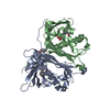
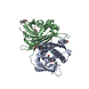
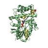
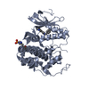
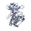
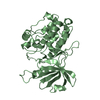
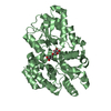
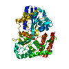
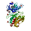
| Title | Hydroxynitrile lyase from the fern Davallia tyermanii | ||||||
 Components Components | Hydroxynitrile lyase | ||||||
 Keywords Keywords |  LYASE / hydroxynitrile lyase / fern LYASE / hydroxynitrile lyase / fern | ||||||
| Function / homology | START domain / Alpha-D-Glucose-1,6-Bisphosphate; Chain A, domain 4 / START-like domain superfamily / lyase activity / 2-Layer Sandwich / Alpha Beta / Hydroxynitrile lyase Function and homology information Function and homology information | ||||||
| Biological species |  Davallia tyermannii (plant) Davallia tyermannii (plant) | ||||||
| Method | X-RAY DIFFRACTION / SYNCHROTRON / SAD / Resolution: 1.854 Å | ||||||
 Authors Authors | Pavkov-Keller, T. / Diepold, M. / Gruber, K. | ||||||
| Funding support |  Austria, 1items Austria, 1items
| ||||||
 Citation Citation | Journal: Sci Rep / Year: 2017 Title: Enzyme discovery beyond homology: a unique hydroxynitrile lyase in the Bet v1 superfamily. Authors: Lanfranchi, E. / Pavkov-Keller, T. / Koehler, E.M. / Diepold, M. / Steiner, K. / Darnhofer, B. / Hartler, J. / Van Den Bergh, T. / Joosten, H.J. / Gruber-Khadjawi, M. / Thallinger, G.G. / ...Authors: Lanfranchi, E. / Pavkov-Keller, T. / Koehler, E.M. / Diepold, M. / Steiner, K. / Darnhofer, B. / Hartler, J. / Van Den Bergh, T. / Joosten, H.J. / Gruber-Khadjawi, M. / Thallinger, G.G. / Birner-Gruenberger, R. / Gruber, K. / Winkler, M. / Glieder, A. | ||||||
| History |
|
- Structure visualization
| Structure viewer | Molecule: MolmilJmol/JSmol |
|---|
- Downloads & links
-Download
| PDBx/mmCIF format | 5e46.cif.gz | 92.9 KB | Display | PDBx/mmCIF format |
|---|---|---|---|---|
| PDB format | pdb5e46.ent.gz | 74.9 KB | Display | PDB format |
| PDBx/mmJSON format | 5e46.json.gz | Tree view | PDBx/mmJSON format | |
| Others |  Other downloads Other downloads |
-Validation report
| Arichive directory | https://data.pdbj.org/pub/pdb/validation_reports/e4/5e46ftp://data.pdbj.org/pub/pdb/validation_reports/e4/5e46 | HTTPS FTP |
|---|
-Related structure data


-Links
 PDBj
PDBj- Assembly
| Deposited unit | 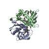
| ||||||||
|---|---|---|---|---|---|---|---|---|---|
| 1 |
| ||||||||
| Unit cell |
|
-Components
| #1: Protein | Mass: 23541.344 Da / Num. of mol.: 2 Source method: isolated from a genetically manipulated source Details: GenBank asseccion number KT804569 / Source: (gene. exp.) Davallia tyermannii (plant) / Production host:  Escherichia coli (E. coli) / References: UniProt: A0A1C9V3S9*PLUS Escherichia coli (E. coli) / References: UniProt: A0A1C9V3S9*PLUS#2: Water | ChemComp-HOH / | Water Mass: 18.015 Da / Num. of mol.: 467 / Source method: isolated from a natural source / Formula: H2O Mass: 18.015 Da / Num. of mol.: 467 / Source method: isolated from a natural source / Formula: H2O |
|---|
-Experimental details
-Experiment
| Experiment | Method: X-RAY DIFFRACTION |
|---|
- Sample preparation
| Crystal | Density Matthews: 2.15 Å3/Da / Density % sol: 42.83 % |
|---|---|
| Crystal grow | Temperature: 298 K / Method: vapor diffusion, sitting drop / pH: 8.5 Details: Native crystals of DtHNL1 were obtained by mixing 0.5ul 4 mg/mL protein sample (in 10 mM Tris-HCl pH 8.0) with 1 ul reservoir solution (0.9 M NaNO3; Na2HPO4; (NH4)2SO4 mix, 0.1 M Tris-Bicine ...Details: Native crystals of DtHNL1 were obtained by mixing 0.5ul 4 mg/mL protein sample (in 10 mM Tris-HCl pH 8.0) with 1 ul reservoir solution (0.9 M NaNO3; Na2HPO4; (NH4)2SO4 mix, 0.1 M Tris-Bicine Buffer pH 8.5 and 30% (w/v) polyethylene glycol monomethyl ether 550 & polyethylene glycol 20k; Morpheus condition C9). Additionally, native crystals were also grown by mixing 1 ul 4 mg/mL protein sample (in 10 mM Tris-HCl pH 8.0) with 0.5ul reservoir solution (0.1 M 2-(4-(2-hydroxyethyl)-1-piperazinyl) ethanesulfonic acid pH 7.5 and 10% (w/v) polyethylene glycol; JSCG condition B4). SeMet-DtHNL1 crystals were obtained in 0.2 M sodium thiocyanate, 20% (w/v) polyethylene glycol 3350. A 1:1 ratio of protein and screening solutions was used, using protein concentration of 3 mg/mL (in 10 mM Tris-HCl pH 8.0). |
-Data collection
| Diffraction | Mean temperature: 100 K |
|---|---|
| Diffraction source | Source: SYNCHROTRON / Site: ESRF  / Beamline: ID29 / Wavelength: 0.979 Å / Beamline: ID29 / Wavelength: 0.979 Å |
| Detector | Type: DECTRIS PILATUS 6M / Detector: PIXEL / Date: May 9, 2014 |
| Radiation | Protocol: SINGLE WAVELENGTH / Monochromatic (M) / Laue (L): M / Scattering type: x-ray |
| Radiation wavelength | Wavelength: 0.979 Å / Relative weight: 1 |
| Reflection | Resolution: 1.85→58 Å / Num. obs: 34750 / % possible obs: 99.84 % / Redundancy: 13.3 % / Rmerge(I) obs: 0.188 / Net I/σ(I): 9.97 |
| Reflection shell | Resolution: 1.85→1.92 Å / Redundancy: 13 % / Rmerge(I) obs: 0.796 / Mean I/σ(I) obs: 2.57 / % possible all: 98.6 |
- Processing
| Software |
| |||||||||||||||||||||||||||||||||||||||||||||||||||||||||||||||||||||||||||||||||||||||||||
|---|---|---|---|---|---|---|---|---|---|---|---|---|---|---|---|---|---|---|---|---|---|---|---|---|---|---|---|---|---|---|---|---|---|---|---|---|---|---|---|---|---|---|---|---|---|---|---|---|---|---|---|---|---|---|---|---|---|---|---|---|---|---|---|---|---|---|---|---|---|---|---|---|---|---|---|---|---|---|---|---|---|---|---|---|---|---|---|---|---|---|---|---|
| Refinement | Method to determine structure: SAD / Resolution: 1.854→57.971 Å / SU ML: 0.16 / Cross valid method: FREE R-VALUE / σ(F): 1.38 / Phase error: 18.1 / Stereochemistry target values: ML
| |||||||||||||||||||||||||||||||||||||||||||||||||||||||||||||||||||||||||||||||||||||||||||
| Solvent computation | Shrinkage radii: 0.9 Å / VDW probe radii: 1.11 Å / Solvent model: FLAT BULK SOLVENT MODEL | |||||||||||||||||||||||||||||||||||||||||||||||||||||||||||||||||||||||||||||||||||||||||||
| Refinement step | Cycle: LAST / Resolution: 1.854→57.971 Å
| |||||||||||||||||||||||||||||||||||||||||||||||||||||||||||||||||||||||||||||||||||||||||||
| Refine LS restraints |
| |||||||||||||||||||||||||||||||||||||||||||||||||||||||||||||||||||||||||||||||||||||||||||
| LS refinement shell |
|
