 Movie
Movie Controller


+ Open data

- Basic information
| Entry | Database: PDB / ID: 2w8b | ||||||
|---|---|---|---|---|---|---|---|
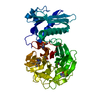
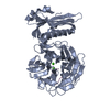
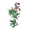
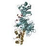
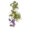
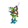
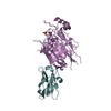
| Title | Crystal structure of processed TolB in complex with Pal | ||||||
 Components Components |
| ||||||
 Keywords Keywords | PROTEIN TRANSPORT/MEMBRANE PROTEIN / PROTEIN TRANSPORT MEMBRANE PROTEIN COMPLEX / TOL / PAL /  TOLB / MEMBRANE / PALMITATE / PERIPLASM / BACTERIOCIN TRANSPORT / TRANSPORT PROTEIN/LIPOPROTEIN / CELL OUTER MEMBRANE / TRANSPORT / LIPOPROTEIN / CELL MEMBRANE / OUTER MEMBRANE / PROTEIN TRANSPORT-MEMBRANE PROTEIN complex TOLB / MEMBRANE / PALMITATE / PERIPLASM / BACTERIOCIN TRANSPORT / TRANSPORT PROTEIN/LIPOPROTEIN / CELL OUTER MEMBRANE / TRANSPORT / LIPOPROTEIN / CELL MEMBRANE / OUTER MEMBRANE / PROTEIN TRANSPORT-MEMBRANE PROTEIN complex | ||||||
| Function / homology |  Function and homology information Function and homology informationcellular response to bacteriocin / regulation of membrane invagination / bacteriocin transport / protein import / cell division site / cell outer membrane / protein transport / outer membrane-bounded periplasmic space / periplasmic space / cell cycle ...cellular response to bacteriocin / regulation of membrane invagination / bacteriocin transport / protein import / cell division site / cell outer membrane / protein transport / outer membrane-bounded periplasmic space / periplasmic space / cell cycle / cell division / protein domain specific binding / protein-containing complex binding / protein-containing complex / membraneSimilarity search - Function | ||||||
| Biological species |  ESCHERICHIA COLI (E. coli) ESCHERICHIA COLI (E. coli) | ||||||
| Method | X-RAY DIFFRACTION / SYNCHROTRON / MOLECULAR REPLACEMENT / Resolution: 1.86 Å | ||||||
 Authors Authors | Sharma, A. / Bonsor, D.A. / Kleanthous, C. | ||||||
 Citation Citation | Journal: Embo J. / Year: 2009 Title: Allosteric Beta-Propeller Signalling in Tolb and its Manipulation by Translocating Colicins. Authors: Bonsor, D.A. / Hecht, O. / Vankemmelbeke, M. / Sharma, A. / Krachler, A.M. / Housden, N.G. / Lilly, K.J. / James, R. / Moore, G.R. / Kleanthous, C. | ||||||
| History |
| ||||||
| Remark 700 | SHEET THE SHEET STRUCTURE OF THIS MOLECULE IS BIFURCATED. IN ORDER TO REPRESENT THIS FEATURE IN ... SHEET THE SHEET STRUCTURE OF THIS MOLECULE IS BIFURCATED. IN ORDER TO REPRESENT THIS FEATURE IN THE SHEET RECORDS BELOW, TWO SHEETS ARE DEFINED. |
- Structure visualization
| Structure viewer | Molecule: MolmilJmol/JSmol |
|---|
- Downloads & links
-Download
| PDBx/mmCIF format | 2w8b.cif.gz | 429.6 KB | Display | PDBx/mmCIF format |
|---|---|---|---|---|
| PDB format | pdb2w8b.ent.gz | 349.3 KB | Display | PDB format |
| PDBx/mmJSON format | 2w8b.json.gz | Tree view | PDBx/mmJSON format | |
| Others |  Other downloads Other downloads |
-Validation report
| Arichive directory | https://data.pdbj.org/pub/pdb/validation_reports/w8/2w8bftp://data.pdbj.org/pub/pdb/validation_reports/w8/2w8b | HTTPS FTP |
|---|
-Related structure data
| Related structure data | 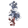 2hqsS S: Starting model for refinement |
|---|---|
| Similar structure data |


-Links
 PDBj
PDBj


- Assembly
| Deposited unit | 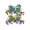
| ||||||||
|---|---|---|---|---|---|---|---|---|---|
| 1 | 
| ||||||||
| 2 | 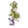
| ||||||||
| 3 | 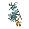
| ||||||||
| 4 | 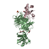
| ||||||||
| Unit cell |
|
-Components
-Protein , 3 types, 8 molecules ABDFCEGH
| #1: Protein | Mass: 43637.379 Da / Num. of mol.: 1 Source method: isolated from a genetically manipulated source Source: (gene. exp.) ESCHERICHIA COLI (E. coli) / Production host: ESCHERICHIA COLI (E. coli) / Strain (production host): BL21(DE3) / References: UniProt: P0A855 | ||
|---|---|---|---|
| #2: Protein | Mass: 43637.379 Da / Num. of mol.: 3 Source method: isolated from a genetically manipulated source Source: (gene. exp.) ESCHERICHIA COLI (E. coli) / Production host: ESCHERICHIA COLI (E. coli) / Strain (production host): BL21(DE3) / References: UniProt: P0A855#3: Protein | Mass: 13518.095 Da / Num. of mol.: 4 / Fragment: RESIDUES 65-173 Source method: isolated from a genetically manipulated source Source: (gene. exp.) ESCHERICHIA COLI (E. coli) / Production host: ESCHERICHIA COLI (E. coli) / Strain (production host): BL21(DE3) / References: UniProt: P0A912 |
-Non-polymers , 4 types, 1538 molecules 


| #4: Chemical | ChemComp-SO4 / Sulfate Mass: 96.063 Da / Num. of mol.: 8 / Source method: obtained synthetically / Formula: SO4 Mass: 96.063 Da / Num. of mol.: 8 / Source method: obtained synthetically / Formula: SO4#5: Chemical | ChemComp-GOL / Glycerol Mass: 92.094 Da / Num. of mol.: 7 / Source method: obtained synthetically / Formula: C3H8O3 Mass: 92.094 Da / Num. of mol.: 7 / Source method: obtained synthetically / Formula: C3H8O3#6: Chemical | ChemComp-ACT / Acetate Mass: 59.044 Da / Num. of mol.: 4 / Source method: obtained synthetically / Formula: C2H3O2 Mass: 59.044 Da / Num. of mol.: 4 / Source method: obtained synthetically / Formula: C2H3O2#7: Water | ChemComp-HOH / | WaterMass: 18.015 Da / Num. of mol.: 1519 / Source method: isolated from a natural source / Formula: H2O |
|---|
-Experimental details
-Experiment
| Experiment | Method: X-RAY DIFFRACTION / Number of used crystals: 1 |
|---|
- Sample preparation
| Crystal | Density Matthews: 2.35 Å3/Da / Density % sol: 47.69 % / Description: NONE |
|---|---|
| Crystal grow | pH: 4.6 Details: 0.1M SODIUM ACETATE PH 4.6, 17% PEG4000, 0.2M AMMONIUM SULPHATE, 20MG/ML |
-Data collection
| Diffraction | Mean temperature: 100 K |
|---|---|
| Diffraction source | Source: SYNCHROTRON / Site: ESRF  / Beamline: ID23-1 / Wavelength: 0.98 / Beamline: ID23-1 / Wavelength: 0.98 |
| Detector | Type: ADSC CCD / Detector: CCD / Date: Mar 8, 2008 |
| Radiation | Protocol: SINGLE WAVELENGTH / Monochromatic (M) / Laue (L): M / Scattering type: x-ray |
| Radiation wavelength | Wavelength: 0.98 Å / Relative weight: 1 |
| Reflection | Resolution: 1.86→45.74 Å / Num. obs: 176410 / % possible obs: 96.2 % / Observed criterion σ(I): 2 / Redundancy: 2.1 % / Rmerge(I) obs: 0.11 / Net I/σ(I): 7.2 |
| Reflection shell | Resolution: 1.86→1.96 Å / Redundancy: 2.1 % / Rmerge(I) obs: 0.66 / Mean I/σ(I) obs: 1.2 / % possible all: 94.9 |
- Processing
| Software |
| ||||||||||||||||||||||||||||||||||||||||||||||||||||||||||||||||||||||||||||||||||||||||||||||||||||||||||||||||||||||||||||||||||||||||||||||||||||||||||||||||||||||||||||||||||||||
|---|---|---|---|---|---|---|---|---|---|---|---|---|---|---|---|---|---|---|---|---|---|---|---|---|---|---|---|---|---|---|---|---|---|---|---|---|---|---|---|---|---|---|---|---|---|---|---|---|---|---|---|---|---|---|---|---|---|---|---|---|---|---|---|---|---|---|---|---|---|---|---|---|---|---|---|---|---|---|---|---|---|---|---|---|---|---|---|---|---|---|---|---|---|---|---|---|---|---|---|---|---|---|---|---|---|---|---|---|---|---|---|---|---|---|---|---|---|---|---|---|---|---|---|---|---|---|---|---|---|---|---|---|---|---|---|---|---|---|---|---|---|---|---|---|---|---|---|---|---|---|---|---|---|---|---|---|---|---|---|---|---|---|---|---|---|---|---|---|---|---|---|---|---|---|---|---|---|---|---|---|---|---|---|
| Refinement | Method to determine structure: MOLECULAR REPLACEMENT Starting model: PDB ENTRY 2HQS Resolution: 1.86→45.36 Å / Cor.coef. Fo:Fc: 0.954 / Cor.coef. Fo:Fc free: 0.924 / SU B: 3.693 / SU ML: 0.108 / Cross valid method: THROUGHOUT / ESU R: 0.155 / ESU R Free: 0.153 / Stereochemistry target values: MAXIMUM LIKELIHOOD / Details: HYDROGENS HAVE BEEN ADDED IN THE RIDING POSITIONS.
| ||||||||||||||||||||||||||||||||||||||||||||||||||||||||||||||||||||||||||||||||||||||||||||||||||||||||||||||||||||||||||||||||||||||||||||||||||||||||||||||||||||||||||||||||||||||
| Solvent computation | Ion probe radii: 0.8 Å / Shrinkage radii: 0.8 Å / VDW probe radii: 1.2 Å / Solvent model: MASK | ||||||||||||||||||||||||||||||||||||||||||||||||||||||||||||||||||||||||||||||||||||||||||||||||||||||||||||||||||||||||||||||||||||||||||||||||||||||||||||||||||||||||||||||||||||||
| Displacement parameters | Biso mean: 19.79 Å2
| ||||||||||||||||||||||||||||||||||||||||||||||||||||||||||||||||||||||||||||||||||||||||||||||||||||||||||||||||||||||||||||||||||||||||||||||||||||||||||||||||||||||||||||||||||||||
| Refinement step | Cycle: LAST / Resolution: 1.86→45.36 Å
| ||||||||||||||||||||||||||||||||||||||||||||||||||||||||||||||||||||||||||||||||||||||||||||||||||||||||||||||||||||||||||||||||||||||||||||||||||||||||||||||||||||||||||||||||||||||
| Refine LS restraints |
|