 Movie
Movie Controller


+ Open data

- Basic information
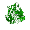
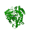
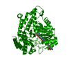
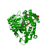
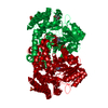
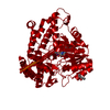
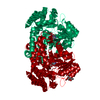
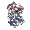
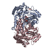
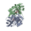
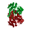
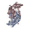
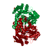
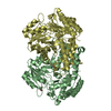
| Entry | Database: PDB / ID: 2w7f | ||||||
|---|---|---|---|---|---|---|---|
| Title | Crystal structure of Y51FbsSHMT L-Ser external aldimine | ||||||
 Components Components | SERINE HYDROXYMETHYLTRANSFERASE | ||||||
 Keywords Keywords | TRANSFERASE / ONE-CARBON METABOLISM / PLP-DEPENDENT ENZYMES | ||||||
| Function / homology |  Function and homology informationglycine hydroxymethyltransferase / glycine hydroxymethyltransferase activity / glycine biosynthetic process from serine / tetrahydrofolate interconversion / pyridoxal phosphate binding / cytoplasm Function and homology informationglycine hydroxymethyltransferase / glycine hydroxymethyltransferase activity / glycine biosynthetic process from serine / tetrahydrofolate interconversion / pyridoxal phosphate binding / cytoplasmSimilarity search - Function | ||||||
| Biological species |   GEOBACILLUS STEAROTHERMOPHILUS (bacteria) GEOBACILLUS STEAROTHERMOPHILUS (bacteria) | ||||||
| Method | X-RAY DIFFRACTION / MOLECULAR REPLACEMENT / Resolution: 1.67 Å | ||||||
 Authors Authors | Rajaram, V. / Bhavani, B.S. / Bisht, S. / Kaul, P. / Prakash, V. / Appaji Rao, N. / Savithri, H.S. / Murthy, M.R.N. | ||||||
 Citation Citation | Journal: FEBS J. / Year: 2008 Title: Importance of Tyrosine Residues of Bacillus Stearothermophilus Serine Hydroxymethyltransferase in Cofactor Binding and L-Allo-Thr Cleavage. Authors: Bhavani, B.S. / Rajaram, V. / Bisht, S. / Kaul, P. / Prakash, V. / Murthy, M.R.N. / Appaji Rao, N. / Savithri, H.S. | ||||||
| History |
|
- Structure visualization
| Structure viewer | Molecule: MolmilJmol/JSmol |
|---|
- Downloads & links
-Download
| PDBx/mmCIF format | 2w7f.cif.gz | 102.3 KB | Display | PDBx/mmCIF format |
|---|---|---|---|---|
| PDB format | pdb2w7f.ent.gz | 77.2 KB | Display | PDB format |
| PDBx/mmJSON format | 2w7f.json.gz | Tree view | PDBx/mmJSON format | |
| Others |  Other downloads Other downloads |
-Validation report
| Arichive directory | https://data.pdbj.org/pub/pdb/validation_reports/w7/2w7fftp://data.pdbj.org/pub/pdb/validation_reports/w7/2w7f | HTTPS FTP |
|---|
-Related structure data
| Related structure data |  2w7dC 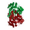 2w7eC  2w7gC  2w7hC 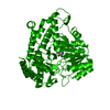 2w7iC 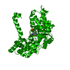 2w7jC 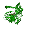 2w7kC  2w7lC  2w7mC  1kl1S S: Starting model for refinement C: citing same article ( |
|---|---|
| Similar structure data |


-Links
 PDBj
PDBj- Assembly
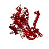
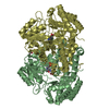
| Deposited unit | 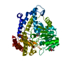
| ||||||||
|---|---|---|---|---|---|---|---|---|---|
| 1 | 
| ||||||||
| Unit cell |
|
-Components
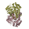
-Protein , 1 types, 1 molecules A
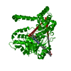
| #1: Protein | Mass: 44258.184 Da / Num. of mol.: 1 / Fragment: RESIDUES 1-405 / Mutation: YES Source method: isolated from a genetically manipulated source Details: SCHIFF LINKAGE BETWEEN LYS A226 AND PLP A501 Source: (gene. exp.) GEOBACILLUS STEAROTHERMOPHILUS (bacteria)Production host: ESCHERICHIA COLI (E. coli) / Strain (production host): BL21References: UniProt: Q7SIB6, glycine hydroxymethyltransferase |
|---|
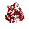
-Non-polymers , 5 types, 440 molecules 


| #2: Chemical | ChemComp-SER / Serine Type: L-peptide linking / Mass: 105.093 Da / Num. of mol.: 1 / Source method: obtained synthetically / Formula: C3H7NO3 Type: L-peptide linking / Mass: 105.093 Da / Num. of mol.: 1 / Source method: obtained synthetically / Formula: C3H7NO3 | ||||
|---|---|---|---|---|---|
| #3: Chemical | ChemComp-PLP / Pyridoxal phosphate Mass: 247.142 Da / Num. of mol.: 1 / Source method: obtained synthetically / Formula: C8H10NO6P Mass: 247.142 Da / Num. of mol.: 1 / Source method: obtained synthetically / Formula: C8H10NO6P | ||||
| #4: Chemical | 2-Methyl-2,4-pentanediol Mass: 118.174 Da / Num. of mol.: 2 / Source method: obtained synthetically / Formula: C6H14O2 / Comment: precipitant*YM Mass: 118.174 Da / Num. of mol.: 2 / Source method: obtained synthetically / Formula: C6H14O2 / Comment: precipitant*YM#5: Chemical | ChemComp-PO4 / | Phosphate Mass: 94.971 Da / Num. of mol.: 1 / Source method: obtained synthetically / Formula: PO4 Mass: 94.971 Da / Num. of mol.: 1 / Source method: obtained synthetically / Formula: PO4#6: Water | ChemComp-HOH / | WaterMass: 18.015 Da / Num. of mol.: 435 / Source method: isolated from a natural source / Formula: H2O |
-Details
| Compound details | ENGINEERED |
|---|
-Experimental details
-Experiment
| Experiment | Method: X-RAY DIFFRACTION / Number of used crystals: 1 |
|---|
- Sample preparation
| Crystal | Density Matthews: 2.1 Å3/Da / Density % sol: 41 % / Description: NONE |
|---|---|
| Crystal grow | Details: 50% MPD, 0.1 M HEPES PH 7.5, 0.2 MM EDTA, 5 MM 2-MERCAPTOETHANOL, 10 MM L-SER |
-Data collection
| Diffraction | Mean temperature: 100 K |
|---|---|
| Diffraction source | Source: ROTATING ANODE / Type: RIGAKU RU200 / Wavelength: 1.5418 |
| Detector | Type: MARRESEARCH / Detector: IMAGE PLATE / Details: OSMIC MIRRORS |
| Radiation | Monochromator: NI FILTER / Protocol: SINGLE WAVELENGTH / Monochromatic (M) / Laue (L): M / Scattering type: x-ray |
| Radiation wavelength | Wavelength: 1.5418 Å / Relative weight: 1 |
| Reflection | Resolution: 1.66→30 Å / Num. obs: 43037 / % possible obs: 96.5 % / Observed criterion σ(I): 0 / Redundancy: 20.1 % / Biso Wilson estimate: 23.3 Å2 / Rmerge(I) obs: 0.06 / Net I/σ(I): 24.8 |
| Reflection shell | Resolution: 1.66→1.72 Å / Rmerge(I) obs: 0.45 / Mean I/σ(I) obs: 3.8 / % possible all: 83.7 |
- Processing
| Software |
| ||||||||||||||||||||||||||||||||||||||||||||||||||||||||||||||||||||||||||||||||||||||||||||||||||||||||||||||||||||||||||||||||||||||||||||||||||||||||||||||||||||||||||||||||||||||
|---|---|---|---|---|---|---|---|---|---|---|---|---|---|---|---|---|---|---|---|---|---|---|---|---|---|---|---|---|---|---|---|---|---|---|---|---|---|---|---|---|---|---|---|---|---|---|---|---|---|---|---|---|---|---|---|---|---|---|---|---|---|---|---|---|---|---|---|---|---|---|---|---|---|---|---|---|---|---|---|---|---|---|---|---|---|---|---|---|---|---|---|---|---|---|---|---|---|---|---|---|---|---|---|---|---|---|---|---|---|---|---|---|---|---|---|---|---|---|---|---|---|---|---|---|---|---|---|---|---|---|---|---|---|---|---|---|---|---|---|---|---|---|---|---|---|---|---|---|---|---|---|---|---|---|---|---|---|---|---|---|---|---|---|---|---|---|---|---|---|---|---|---|---|---|---|---|---|---|---|---|---|---|---|
| Refinement | Method to determine structure: MOLECULAR REPLACEMENT Starting model: PDB ENTRY 1KL1 Resolution: 1.67→22.45 Å / Cor.coef. Fo:Fc: 0.96 / Cor.coef. Fo:Fc free: 0.945 / SU B: 1.882 / SU ML: 0.065 / Cross valid method: THROUGHOUT / ESU R: 0.114 / ESU R Free: 0.108 / Stereochemistry target values: MAXIMUM LIKELIHOOD / Details: HYDROGENS HAVE BEEN ADDED IN THE RIDING POSITIONS.
| ||||||||||||||||||||||||||||||||||||||||||||||||||||||||||||||||||||||||||||||||||||||||||||||||||||||||||||||||||||||||||||||||||||||||||||||||||||||||||||||||||||||||||||||||||||||
| Solvent computation | Ion probe radii: 0.8 Å / Shrinkage radii: 0.8 Å / VDW probe radii: 1.4 Å / Solvent model: MASK | ||||||||||||||||||||||||||||||||||||||||||||||||||||||||||||||||||||||||||||||||||||||||||||||||||||||||||||||||||||||||||||||||||||||||||||||||||||||||||||||||||||||||||||||||||||||
| Displacement parameters | Biso mean: 19.01 Å2
| ||||||||||||||||||||||||||||||||||||||||||||||||||||||||||||||||||||||||||||||||||||||||||||||||||||||||||||||||||||||||||||||||||||||||||||||||||||||||||||||||||||||||||||||||||||||
| Refinement step | Cycle: LAST / Resolution: 1.67→22.45 Å
| ||||||||||||||||||||||||||||||||||||||||||||||||||||||||||||||||||||||||||||||||||||||||||||||||||||||||||||||||||||||||||||||||||||||||||||||||||||||||||||||||||||||||||||||||||||||
| Refine LS restraints |
|