 Movie
Movie Controller

[English] 日本語
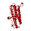
 Yorodumi
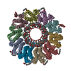
- PDB-2vty: Vaccinia virus anti-apoptotic F1L is a novel Bcl-2-like domain sw... -
+ Open data

- Basic information
| Entry | Database: PDB / ID: 2vty | ||||||
|---|---|---|---|---|---|---|---|
| Title | Vaccinia virus anti-apoptotic F1L is a novel Bcl-2-like domain swapped dimer | ||||||
 Components Components | PROTEIN F1 | ||||||
 Keywords Keywords |  APOPTOSIS / BCL-2 / VACCINIA VIRUS APOPTOSIS / BCL-2 / VACCINIA VIRUS | ||||||
| Function / homology |  Function and homology information Function and homology information: / host cell mitochondrial outer membrane / : / regulation of apoptotic process / membraneSimilarity search - Function | ||||||
| Biological species |   VACCINIA VIRUS VACCINIA VIRUS | ||||||
| Method | X-RAY DIFFRACTION / SIRAS / Resolution: 2.1 Å | ||||||
 Authors Authors | Kvansakul, M. / Yang, H. / Fairlie, W.D. / Czabotar, P.E. / Fischer, S.F. / Perugini, M.A. / Huang, D.C.S. / Colman, P.M. | ||||||
 Citation Citation | Journal: Cell Death Differ. / Year: 2008 Title: Vaccinia Virus Anti-Apoptotic F1L is a Novel Bcl-2-Like Domain-Swapped Dimer that Binds a Highly Selective Subset of Bh3-Containing Death Ligands. Authors: Kvansakul, M. / Yang, H. / Fairlie, W.D. / Czabotar, P.E. / Fischer, S.F. / Perugini, M.A. / Huang, D.C.S. / Colman, P.M. | ||||||
| History |
|
- Structure visualization
| Structure viewer | Molecule: MolmilJmol/JSmol |
|---|
- Downloads & links
-Download
| PDBx/mmCIF format | 2vty.cif.gz | 43.5 KB | Display | PDBx/mmCIF format |
|---|---|---|---|---|
| PDB format | pdb2vty.ent.gz | 30.9 KB | Display | PDB format |
| PDBx/mmJSON format | 2vty.json.gz | Tree view | PDBx/mmJSON format | |
| Others |  Other downloads Other downloads |
-Validation report
| Arichive directory | https://data.pdbj.org/pub/pdb/validation_reports/vt/2vtyftp://data.pdbj.org/pub/pdb/validation_reports/vt/2vty | HTTPS FTP |
|---|
-Related structure data
| Similar structure data |
|---|

-Links
 PDBj
PDBj
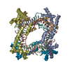
- Assembly
| Deposited unit | 
| ||||||||
|---|---|---|---|---|---|---|---|---|---|
| 1 | x 12
| ||||||||
| Unit cell |
|
-Components
| #1: Protein | Mass: 21456.246 Da / Num. of mol.: 1 / Fragment: RESIDUES 18-186 Source method: isolated from a genetically manipulated source Source: (gene. exp.) VACCINIA VIRUS / Strain: MODIFIED VACCINIA ANKARA / Plasmid: PET DUET / Production host:  ESCHERICHIA COLI (E. coli) / Strain (production host): BL21(DE3) PLYSS / References: UniProt: O57173 ESCHERICHIA COLI (E. coli) / Strain (production host): BL21(DE3) PLYSS / References: UniProt: O57173 |
|---|---|
| #2: Water | ChemComp-HOH / Water Mass: 18.015 Da / Num. of mol.: 66 / Source method: isolated from a natural source / Formula: H2O Mass: 18.015 Da / Num. of mol.: 66 / Source method: isolated from a natural source / Formula: H2O |
| Sequence details | N-TERMINAL HEXAHISTID |
-Experimental details
-Experiment
| Experiment | Method: X-RAY DIFFRACTION / Number of used crystals: 1 |
|---|
- Sample preparation
| Crystal | Density Matthews: 2.67 Å3/Da / Density % sol: 54 % / Description: NONE |
|---|---|
| Crystal grow | pH: 5.2 / Details: 0.9 NACL, 50 MM NA/K PHOSPHATE PH 5.2 |
-Data collection
| Diffraction | Mean temperature: 100 K |
|---|---|
| Diffraction source | Source: ROTATING ANODE / Type: RIGAKU MICROMAX-007 / Wavelength: 1.5418 |
| Detector | Type: RIGAKU IMAGE PLATE / Detector: IMAGE PLATE / Date: Aug 10, 2006 / Details: CAPILLARY OPTICS |
| Radiation | Protocol: SINGLE WAVELENGTH / Monochromatic (M) / Laue (L): M / Scattering type: x-ray |
| Radiation wavelength | Wavelength: 1.5418 Å / Relative weight: 1 |
| Reflection | Resolution: 2.1→50 Å / Num. obs: 13687 / % possible obs: 99.4 % / Observed criterion σ(I): 2 / Redundancy: 5.1 % / Rmerge(I) obs: 0.09 / Net I/σ(I): 14.4 |
| Reflection shell | Resolution: 2.1→2.18 Å / Redundancy: 5.1 % / Rmerge(I) obs: 0.55 / Mean I/σ(I) obs: 3.5 / % possible all: 99.9 |
- Processing
| Software |
| ||||||||||||||||||||||||||||||||||||||||||||||||||||||||||||||||||||||||||||||||||||||||||||||||||||||||||||||||||||||||||||||||||||||||||||||||||||||||||||||||||||||||||||||||||||||
|---|---|---|---|---|---|---|---|---|---|---|---|---|---|---|---|---|---|---|---|---|---|---|---|---|---|---|---|---|---|---|---|---|---|---|---|---|---|---|---|---|---|---|---|---|---|---|---|---|---|---|---|---|---|---|---|---|---|---|---|---|---|---|---|---|---|---|---|---|---|---|---|---|---|---|---|---|---|---|---|---|---|---|---|---|---|---|---|---|---|---|---|---|---|---|---|---|---|---|---|---|---|---|---|---|---|---|---|---|---|---|---|---|---|---|---|---|---|---|---|---|---|---|---|---|---|---|---|---|---|---|---|---|---|---|---|---|---|---|---|---|---|---|---|---|---|---|---|---|---|---|---|---|---|---|---|---|---|---|---|---|---|---|---|---|---|---|---|---|---|---|---|---|---|---|---|---|---|---|---|---|---|---|---|
| Refinement | Method to determine structure: SIRAS Starting model: NONE Resolution: 2.1→88.39 Å / Cor.coef. Fo:Fc: 0.943 / Cor.coef. Fo:Fc free: 0.935 / SU B: 3.556 / SU ML: 0.095 / Cross valid method: THROUGHOUT / ESU R: 0.163 / ESU R Free: 0.137 / Stereochemistry target values: MAXIMUM LIKELIHOOD / Details: HYDROGENS HAVE BEEN ADDED IN THE RIDING POSITIONS.
| ||||||||||||||||||||||||||||||||||||||||||||||||||||||||||||||||||||||||||||||||||||||||||||||||||||||||||||||||||||||||||||||||||||||||||||||||||||||||||||||||||||||||||||||||||||||
| Solvent computation | Ion probe radii: 0.8 Å / Shrinkage radii: 0.8 Å / VDW probe radii: 1.4 Å / Solvent model: MASK | ||||||||||||||||||||||||||||||||||||||||||||||||||||||||||||||||||||||||||||||||||||||||||||||||||||||||||||||||||||||||||||||||||||||||||||||||||||||||||||||||||||||||||||||||||||||
| Displacement parameters | Biso mean: 28.971 Å2 | ||||||||||||||||||||||||||||||||||||||||||||||||||||||||||||||||||||||||||||||||||||||||||||||||||||||||||||||||||||||||||||||||||||||||||||||||||||||||||||||||||||||||||||||||||||||
| Refinement step | Cycle: LAST / Resolution: 2.1→88.39 Å
| ||||||||||||||||||||||||||||||||||||||||||||||||||||||||||||||||||||||||||||||||||||||||||||||||||||||||||||||||||||||||||||||||||||||||||||||||||||||||||||||||||||||||||||||||||||||
| Refine LS restraints |
|