 Movie
Movie Controller


+ Open data

- Basic information
| Entry | Database: PDB / ID: 2vsx | ||||||
|---|---|---|---|---|---|---|---|
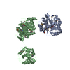
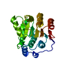
| Title | Crystal Structure of a Translation Initiation Complex | ||||||
 Components Components |
| ||||||
 Keywords Keywords | TRANSLATION/HYDROLASE /  ACETYLATION / ATP-BINDING / PHOSPHOPROTEIN / PROTEIN BIOSYNTHESIS / TRANSLATION REGULATION / TRANSLATION INITIATION / INITIATION FACTOR / NUCLEOTIDE-BINDING / HELICASE / HYDROLASE / CYTOPLASM / RNA-BINDING / TRANSLATION-HYDROLASE complex ACETYLATION / ATP-BINDING / PHOSPHOPROTEIN / PROTEIN BIOSYNTHESIS / TRANSLATION REGULATION / TRANSLATION INITIATION / INITIATION FACTOR / NUCLEOTIDE-BINDING / HELICASE / HYDROLASE / CYTOPLASM / RNA-BINDING / TRANSLATION-HYDROLASE complex | ||||||
| Function / homology |  Function and homology information Function and homology informationDeadenylation of mRNA / Activation of the mRNA upon binding of the cap-binding complex and eIFs, and subsequent binding to 43S / positive regulation of formation of translation preinitiation complex / eukaryotic translation initiation factor 4F complex / cytoplasmic translational initiation / regulation of protein metabolic process / ATP-dependent activity, acting on RNA / positive regulation of endoplasmic reticulum unfolded protein response / mTORC1-mediated signalling / regulation of translational initiation ...Deadenylation of mRNA / Activation of the mRNA upon binding of the cap-binding complex and eIFs, and subsequent binding to 43S / positive regulation of formation of translation preinitiation complex / eukaryotic translation initiation factor 4F complex / cytoplasmic translational initiation / regulation of protein metabolic process / ATP-dependent activity, acting on RNA / positive regulation of endoplasmic reticulum unfolded protein response / mTORC1-mediated signalling / regulation of translational initiation / Translation initiation complex formation / Ribosomal scanning and start codon recognition / ATPase activator activity / Nonsense Mediated Decay (NMD) independent of the Exon Junction Complex (EJC) / Nonsense Mediated Decay (NMD) enhanced by the Exon Junction Complex (EJC) / L13a-mediated translational silencing of Ceruloplasmin expression / stress granule assembly / translational initiation / translation initiation factor activity / ribosomal large subunit biogenesis / molecular condensate scaffold activity / P-body / cytoplasmic stress granule / : / RNA helicase activity / ribosome / RNA helicase / mRNA binding / ATP hydrolysis activity / mitochondrion / RNA binding / ATP binding / plasma membrane / cytoplasmSimilarity search - Function | ||||||
| Biological species |  SACCHAROMYCES CEREVISIAE (brewer's yeast) SACCHAROMYCES CEREVISIAE (brewer's yeast) | ||||||
| Method | X-RAY DIFFRACTION / MOLECULAR REPLACEMENT / Resolution: 2.8 Å | ||||||
 Authors Authors | Schutz, P. / Bumann, M. / Oberholzer, A.E. / Bieniossek, C. / Altmann, M. / Trachsel, H. / Baumann, U. | ||||||
 Citation Citation | Journal: Proc.Natl.Acad.Sci.USA / Year: 2008 Title: Crystal Structure of the Yeast Eif4A-Eif4G Complex: An RNA-Helicase Controlled by Protein-Protein Interactions. Authors: Schutz, P. / Bumann, M. / Oberholzer, A.E. / Bieniossek, C. / Trachsel, H. / Altmann, M. / Baumann, U. | ||||||
| History |
|
- Structure visualization
| Structure viewer | Molecule: MolmilJmol/JSmol |
|---|
- Downloads & links
-Download
| PDBx/mmCIF format | 2vsx.cif.gz | 496.2 KB | Display | PDBx/mmCIF format |
|---|---|---|---|---|
| PDB format | pdb2vsx.ent.gz | 408.7 KB | Display | PDB format |
| PDBx/mmJSON format | 2vsx.json.gz | Tree view | PDBx/mmJSON format | |
| Others |  Other downloads Other downloads |
-Validation report
| Arichive directory | https://data.pdbj.org/pub/pdb/validation_reports/vs/2vsxftp://data.pdbj.org/pub/pdb/validation_reports/vs/2vsx | HTTPS FTP |
|---|
-Related structure data
| Related structure data | 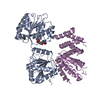 2vsoC 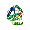 1fukS  1hu3S 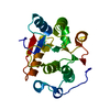 1qdeS S: Starting model for refinement C: citing same article ( |
|---|---|
| Similar structure data |


-Links
 PDBj
PDBj


- Assembly
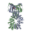
| Deposited unit | 
| |||||||||||||||||||||||||||||||||
|---|---|---|---|---|---|---|---|---|---|---|---|---|---|---|---|---|---|---|---|---|---|---|---|---|---|---|---|---|---|---|---|---|---|---|
| 1 |
| |||||||||||||||||||||||||||||||||
| 2 | 
| |||||||||||||||||||||||||||||||||
| 3 | 
| |||||||||||||||||||||||||||||||||
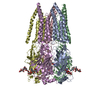
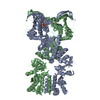
| Unit cell |
| |||||||||||||||||||||||||||||||||
| Noncrystallographic symmetry (NCS) | NCS domain:
NCS domain segments:
NCS ensembles :
|
-Components
| #1: Protein | Mass: 44745.988 Da / Num. of mol.: 2 Source method: isolated from a genetically manipulated source Source: (gene. exp.) SACCHAROMYCES CEREVISIAE (brewer's yeast)Plasmid: PET-28 / Production host:  ESCHERICHIA COLI (E. coli) / Strain (production host): BL21 / Variant (production host): ROSETTA ESCHERICHIA COLI (E. coli) / Strain (production host): BL21 / Variant (production host): ROSETTAReferences: UniProt: P10081, Hydrolases; Acting on acid anhydrides; In phosphorus-containing anhydrides#2: Protein | / EIF4F P150 / EIF-4F P150 / EIF4G1 / MRNA CAP-BINDING PROTEIN COMPLEX SUBUNIT P150 / EIF4GMass: 32360.195 Da / Num. of mol.: 2 / Fragment: MIDDLE DOMAIN, 4A-BINDING, RESIDUES 572-854 Source method: isolated from a genetically manipulated source Source: (gene. exp.) SACCHAROMYCES CEREVISIAE (brewer's yeast)Plasmid: PET-28 / Production host: ESCHERICHIA COLI (E. coli) / Strain (production host): BL21(DE3) / References: UniProt: P39935#3: Chemical | Adenosine monophosphate  Mass: 347.221 Da / Num. of mol.: 2 / Source method: obtained synthetically / Formula: C10H14N5O7P / Comment: AMP*YM Mass: 347.221 Da / Num. of mol.: 2 / Source method: obtained synthetically / Formula: C10H14N5O7P / Comment: AMP*YM |
|---|
-Experimental details
-Experiment
| Experiment | Method: X-RAY DIFFRACTION / Number of used crystals: 1 |
|---|
- Sample preparation
| Crystal | Density Matthews: 3.1 Å3/Da / Density % sol: 60 % / Description: NONE |
|---|---|
| Crystal grow | Temperature: 277 K / pH: 7.2 Details: 0.2 M TARTRATE, 20 % PEG 3350, PH 7.2, 4 DEG C, 10 ,MM AMP |
-Data collection
| Diffraction | Mean temperature: 110 K |
|---|---|
| Diffraction source | Source: ROTATING ANODE / Type: RIGAKU RU300 / Wavelength: 1.5418 |
| Detector | Type: RIGAKU RAXIS-IV / Detector: IMAGE PLATE / Date: Mar 15, 2002 / Details: OSMICS MIRROR |
| Radiation | Monochromator: OSMIC MIRROR / Protocol: SINGLE WAVELENGTH / Monochromatic (M) / Laue (L): M / Scattering type: x-ray |
| Radiation wavelength | Wavelength: 1.5418 Å / Relative weight: 1 |
| Reflection | Resolution: 2.8→50 Å / Num. obs: 40597 / % possible obs: 99.8 % / Observed criterion σ(I): -3 / Redundancy: 3.4 % / Biso Wilson estimate: 37.35 Å2 / Rmerge(I) obs: 0.09 / Net I/σ(I): 11.5 |
| Reflection shell | Resolution: 2.8→2.9 Å / Redundancy: 3 % / Rmerge(I) obs: 0.7 / Mean I/σ(I) obs: 1.6 / % possible all: 98.6 |
- Processing
| Software |
| |||||||||||||||||||||||||||||||||||||||||||||||||||||||||||||||||||||||||||||||||||||||||||||||||||||||||||||||||||||||||||||||||||||||||||||||||||||||||||||||||||||||||||||||||||||||||||||||||||||||||||||||||||||||||||||||||||||||||||||||||||||||||||||||||||||||||||||||||||
|---|---|---|---|---|---|---|---|---|---|---|---|---|---|---|---|---|---|---|---|---|---|---|---|---|---|---|---|---|---|---|---|---|---|---|---|---|---|---|---|---|---|---|---|---|---|---|---|---|---|---|---|---|---|---|---|---|---|---|---|---|---|---|---|---|---|---|---|---|---|---|---|---|---|---|---|---|---|---|---|---|---|---|---|---|---|---|---|---|---|---|---|---|---|---|---|---|---|---|---|---|---|---|---|---|---|---|---|---|---|---|---|---|---|---|---|---|---|---|---|---|---|---|---|---|---|---|---|---|---|---|---|---|---|---|---|---|---|---|---|---|---|---|---|---|---|---|---|---|---|---|---|---|---|---|---|---|---|---|---|---|---|---|---|---|---|---|---|---|---|---|---|---|---|---|---|---|---|---|---|---|---|---|---|---|---|---|---|---|---|---|---|---|---|---|---|---|---|---|---|---|---|---|---|---|---|---|---|---|---|---|---|---|---|---|---|---|---|---|---|---|---|---|---|---|---|---|---|---|---|---|---|---|---|---|---|---|---|---|---|---|---|---|---|---|---|---|---|---|---|---|---|---|---|---|---|---|---|---|---|---|---|---|---|---|---|---|---|---|---|---|---|---|---|---|---|---|
| Refinement | Method to determine structure: MOLECULAR REPLACEMENT Starting model: PDB ENTRIES 1QDE, 1FUK, 1HU3 Resolution: 2.8→33.21 Å / SU ML: 0.41 / Phase error: 35.53 / Stereochemistry target values: ML
| |||||||||||||||||||||||||||||||||||||||||||||||||||||||||||||||||||||||||||||||||||||||||||||||||||||||||||||||||||||||||||||||||||||||||||||||||||||||||||||||||||||||||||||||||||||||||||||||||||||||||||||||||||||||||||||||||||||||||||||||||||||||||||||||||||||||||||||||||||
| Solvent computation | Shrinkage radii: 0.8 Å / VDW probe radii: 1 Å / Solvent model: FLAT BULK SOLVENT MODEL / Bsol: 64.709 Å2 / ksol: 0.334 e/Å3 | |||||||||||||||||||||||||||||||||||||||||||||||||||||||||||||||||||||||||||||||||||||||||||||||||||||||||||||||||||||||||||||||||||||||||||||||||||||||||||||||||||||||||||||||||||||||||||||||||||||||||||||||||||||||||||||||||||||||||||||||||||||||||||||||||||||||||||||||||||
| Displacement parameters | Biso mean: 31.98 Å2
| |||||||||||||||||||||||||||||||||||||||||||||||||||||||||||||||||||||||||||||||||||||||||||||||||||||||||||||||||||||||||||||||||||||||||||||||||||||||||||||||||||||||||||||||||||||||||||||||||||||||||||||||||||||||||||||||||||||||||||||||||||||||||||||||||||||||||||||||||||
| Refinement step | Cycle: LAST / Resolution: 2.8→33.21 Å
| |||||||||||||||||||||||||||||||||||||||||||||||||||||||||||||||||||||||||||||||||||||||||||||||||||||||||||||||||||||||||||||||||||||||||||||||||||||||||||||||||||||||||||||||||||||||||||||||||||||||||||||||||||||||||||||||||||||||||||||||||||||||||||||||||||||||||||||||||||
| Refine LS restraints |
| |||||||||||||||||||||||||||||||||||||||||||||||||||||||||||||||||||||||||||||||||||||||||||||||||||||||||||||||||||||||||||||||||||||||||||||||||||||||||||||||||||||||||||||||||||||||||||||||||||||||||||||||||||||||||||||||||||||||||||||||||||||||||||||||||||||||||||||||||||
| Refine LS restraints NCS |
| |||||||||||||||||||||||||||||||||||||||||||||||||||||||||||||||||||||||||||||||||||||||||||||||||||||||||||||||||||||||||||||||||||||||||||||||||||||||||||||||||||||||||||||||||||||||||||||||||||||||||||||||||||||||||||||||||||||||||||||||||||||||||||||||||||||||||||||||||||
| LS refinement shell |
| |||||||||||||||||||||||||||||||||||||||||||||||||||||||||||||||||||||||||||||||||||||||||||||||||||||||||||||||||||||||||||||||||||||||||||||||||||||||||||||||||||||||||||||||||||||||||||||||||||||||||||||||||||||||||||||||||||||||||||||||||||||||||||||||||||||||||||||||||||
| Refinement TLS params. | Method: refined / Refine-ID: X-RAY DIFFRACTION
| |||||||||||||||||||||||||||||||||||||||||||||||||||||||||||||||||||||||||||||||||||||||||||||||||||||||||||||||||||||||||||||||||||||||||||||||||||||||||||||||||||||||||||||||||||||||||||||||||||||||||||||||||||||||||||||||||||||||||||||||||||||||||||||||||||||||||||||||||||
| Refinement TLS group |
|
