 Movie
Movie Controller


+ Open data

- Basic information
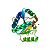
| Entry | Database: PDB / ID: 1fuu | ||||||
|---|---|---|---|---|---|---|---|
| Title | YEAST INITIATION FACTOR 4A | ||||||
 Components Components | YEAST INITIATION FACTOR 4A | ||||||
 Keywords Keywords |  TRANSLATION / IF4A / HELICASE / DEAD-BOX PROTEIN TRANSLATION / IF4A / HELICASE / DEAD-BOX PROTEIN | ||||||
| Function / homology |  Function and homology information Function and homology informationActivation of the mRNA upon binding of the cap-binding complex and eIFs, and subsequent binding to 43S / positive regulation of formation of translation preinitiation complex / eukaryotic translation initiation factor 4F complex / cytoplasmic translational initiation / ATP-dependent activity, acting on RNA / regulation of translational initiation / Translation initiation complex formation / Ribosomal scanning and start codon recognition / L13a-mediated translational silencing of Ceruloplasmin expression / translational initiation ...Activation of the mRNA upon binding of the cap-binding complex and eIFs, and subsequent binding to 43S / positive regulation of formation of translation preinitiation complex / eukaryotic translation initiation factor 4F complex / cytoplasmic translational initiation / ATP-dependent activity, acting on RNA / regulation of translational initiation / Translation initiation complex formation / Ribosomal scanning and start codon recognition / L13a-mediated translational silencing of Ceruloplasmin expression / translational initiation / translation initiation factor activity / cytoplasmic stress granule / RNA helicase activity / ribosome / RNA helicase / ATP hydrolysis activity / RNA binding / ATP binding / plasma membrane / cytoplasmSimilarity search - Function | ||||||
| Biological species |  Saccharomyces cerevisiae (brewer's yeast) Saccharomyces cerevisiae (brewer's yeast) | ||||||
| Method | X-RAY DIFFRACTION / SYNCHROTRON / Resolution: 2.5 Å | ||||||
 Authors Authors | Caruthers, J.M. / Johnson, E.R. / McKay, D.B. | ||||||
 Citation Citation | Journal: Proc.Natl.Acad.Sci.USA / Year: 2000 Title: Crystal structure of yeast initiation factor 4A, a DEAD-box RNA helicase. Authors: Caruthers, J.M. / Johnson, E.R. / McKay, D.B. | ||||||
| History |
|
- Structure visualization
| Structure viewer | Molecule: MolmilJmol/JSmol |
|---|
- Downloads & links
-Download
| PDBx/mmCIF format | 1fuu.cif.gz | 127.8 KB | Display | PDBx/mmCIF format |
|---|---|---|---|---|
| PDB format | pdb1fuu.ent.gz | 105 KB | Display | PDB format |
| PDBx/mmJSON format | 1fuu.json.gz | Tree view | PDBx/mmJSON format | |
| Others |  Other downloads Other downloads |
-Validation report
| Arichive directory | https://data.pdbj.org/pub/pdb/validation_reports/fu/1fuuftp://data.pdbj.org/pub/pdb/validation_reports/fu/1fuu | HTTPS FTP |
|---|
-Related structure data


-Links
 PDBj
PDBj

- Assembly
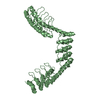
| Deposited unit | 
| ||||||||
|---|---|---|---|---|---|---|---|---|---|
| 1 |
| ||||||||
| Unit cell |
| ||||||||
| Details | the biological assembly is a dumbell of two domains connected by an eleven-residue linker |
-Components
| #1: Protein | Mass: 45130.637 Da / Num. of mol.: 2 / Fragment: MRNA HELICASE Source method: isolated from a genetically manipulated source Source: (gene. exp.) Saccharomyces cerevisiae (brewer's yeast)Production host:  Escherichia coli (E. coli) / References: UniProt: P10081 Escherichia coli (E. coli) / References: UniProt: P10081#2: Water | ChemComp-HOH / | Water Mass: 18.015 Da / Num. of mol.: 146 / Source method: isolated from a natural source / Formula: H2O Mass: 18.015 Da / Num. of mol.: 146 / Source method: isolated from a natural source / Formula: H2O |
|---|
-Experimental details
-Experiment
| Experiment | Method: X-RAY DIFFRACTION / Number of used crystals: 1 |
|---|
- Sample preparation
| Crystal | Density Matthews: 2.19 Å3/Da / Density % sol: 43.92 % | ||||||||||||||||||||||||
|---|---|---|---|---|---|---|---|---|---|---|---|---|---|---|---|---|---|---|---|---|---|---|---|---|---|
| Crystal grow | Temperature: 298 K / Method: vapor diffusion, hanging drop / pH: 9 Details: CHAPS, guanidinium, KOAc, pH 9.0, VAPOR DIFFUSION, HANGING DROP, temperature 298K | ||||||||||||||||||||||||
| Crystal grow | *PLUS pH: 6.5 / Method: unknown | ||||||||||||||||||||||||
| Components of the solutions | *PLUS
|
-Data collection
| Diffraction | Mean temperature: 100 K |
|---|---|
| Diffraction source | Source: SYNCHROTRON / Site: SSRL  / Beamline: BL1-5 / Wavelength: 0.92 / Beamline: BL1-5 / Wavelength: 0.92 |
| Detector | Type: ADSC QUANTUM 4 / Detector: CCD / Date: Jul 20, 1999 |
| Radiation | Protocol: SINGLE WAVELENGTH / Monochromatic (M) / Laue (L): M / Scattering type: x-ray |
| Radiation wavelength | Wavelength: 0.92 Å / Relative weight: 1 |
| Reflection | Resolution: 2.5→40 Å / Num. all: 48114 / Num. obs: 16117 / Observed criterion σ(F): 0 / Observed criterion σ(I): 0 / Redundancy: 2.99 % / Biso Wilson estimate: 20.9 Å2 / Rmerge(I) obs: 0.079 |
| Reflection shell | Highest resolution: 2.5 Å |
| Reflection | *PLUS % possible obs: 92 % / Num. measured all: 53337 / Rmerge(I) obs: 0.071 |
| Reflection shell | *PLUS % possible obs: 80.4 % / Rmerge(I) obs: 0.097 |
- Processing
| Software |
| ||||||||||||||||||||
|---|---|---|---|---|---|---|---|---|---|---|---|---|---|---|---|---|---|---|---|---|---|
| Refinement | Resolution: 2.5→40 Å / σ(F): 0 / σ(I): 0 / Stereochemistry target values: Engh & Huber
| ||||||||||||||||||||
| Refinement step | Cycle: LAST / Resolution: 2.5→40 Å
| ||||||||||||||||||||
| Refine LS restraints |
| ||||||||||||||||||||
| Software | *PLUS Name: CNS / Classification: refinement | ||||||||||||||||||||
| Refinement | *PLUS Rfactor Rfree: 0.273 / Rfactor Rwork: 0.244 | ||||||||||||||||||||
| Solvent computation | *PLUS | ||||||||||||||||||||
| Displacement parameters | *PLUS |
