 Movie
Movie Controller

[English] 日本語
 Yorodumi
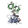
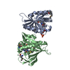
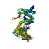
- PDB-2vli: Structure of Deinococcus radiodurans tunicamycin resistance protein -
+ Open data

- Basic information
| Entry | Database: PDB / ID: 2vli | ||||||
|---|---|---|---|---|---|---|---|
| Title | Structure of Deinococcus radiodurans tunicamycin resistance protein | ||||||
 Components Components | ANTIBIOTIC RESISTANCE PROTEIN Antimicrobial resistance Antimicrobial resistance | ||||||
 Keywords Keywords | TRANSFERASE / PHOSPHOTRANSFERASE | ||||||
| Function / homology | P-loop containing nucleotide triphosphate hydrolases / P-loop containing nucleoside triphosphate hydrolase / Rossmann fold / 3-Layer(aba) Sandwich / Alpha Beta / : / Antibiotic resistance protein Function and homology information Function and homology information | ||||||
| Biological species |  DEINOCOCCUS RADIODURANS (radioresistant) DEINOCOCCUS RADIODURANS (radioresistant) | ||||||
| Method | X-RAY DIFFRACTION / SYNCHROTRON / SAD / Resolution: 1.95 Å | ||||||
 Authors Authors | Macedo, S. / Kapp, U. / Leiros, I. / Hall, D.R. / Mitchell, E. | ||||||
 Citation Citation | Journal: Acta Crystallogr.,Sect.F / Year: 2008 Title: Structure of Deinococcus Radiodurans Tunicamycin-Resistance Protein (Tmrd), a Phosphotransferase. Authors: Kapp, U. / Macedo, S. / Hall, D.R. / Leiros, I. / Mcsweeney, S.M. / Mitchell, E. | ||||||
| History |
|
- Structure visualization
| Structure viewer | Molecule: MolmilJmol/JSmol |
|---|
- Downloads & links
-Download
| PDBx/mmCIF format | 2vli.cif.gz | 80 KB | Display | PDBx/mmCIF format |
|---|---|---|---|---|
| PDB format | pdb2vli.ent.gz | 64.5 KB | Display | PDB format |
| PDBx/mmJSON format | 2vli.json.gz | Tree view | PDBx/mmJSON format | |
| Others |  Other downloads Other downloads |
-Validation report
| Arichive directory | https://data.pdbj.org/pub/pdb/validation_reports/vl/2vliftp://data.pdbj.org/pub/pdb/validation_reports/vl/2vli | HTTPS FTP |
|---|
-Related structure data
| Similar structure data |
|---|


-Links
 PDBj
PDBj- Assembly
| Deposited unit | 
| |||||||||
|---|---|---|---|---|---|---|---|---|---|---|
| 1 | 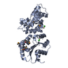
| |||||||||
| 2 | 
| |||||||||
| Unit cell |
| |||||||||
| Components on special symmetry positions |
|
-Components
| #1: Protein | Antimicrobial resistance / TUNICAMYCIN RESISTANCE PROTEIN Mass: 20420.512 Da / Num. of mol.: 2 Source method: isolated from a genetically manipulated source Source: (gene. exp.) DEINOCOCCUS RADIODURANS (radioresistant)Strain: R1 / Production host: ESCHERICHIA COLI (E. coli) / Strain (production host): BL21(DE3) / References: UniProt: Q9RUG7#2: Chemical | ChemComp-CD / |   Mass: 112.411 Da / Num. of mol.: 1 / Source method: obtained synthetically / Formula: Cd Mass: 112.411 Da / Num. of mol.: 1 / Source method: obtained synthetically / Formula: Cd#3: Chemical | ChemComp-CL / | Chloride  Mass: 35.453 Da / Num. of mol.: 1 / Source method: obtained synthetically / Formula: Cl Mass: 35.453 Da / Num. of mol.: 1 / Source method: obtained synthetically / Formula: Cl#4: Water | ChemComp-HOH / | Water Mass: 18.015 Da / Num. of mol.: 226 / Source method: isolated from a natural source / Formula: H2O Mass: 18.015 Da / Num. of mol.: 226 / Source method: isolated from a natural source / Formula: H2ONonpolymer details | CADMIUM ION (CD): FROM CRYSTALLIS | Sequence details | PROTEIN WAS EXPRESSED USING A CONSTRUCT GIVING A PRODUCT WITH A C-TERMINAL TRUNCATION FROM RESIDUE ...PROTEIN WAS EXPRESSED USING A CONSTRUCT GIVING A PRODUCT WITH A C-TERMINAL TRUNCATION | |
|---|
-Experimental details
-Experiment
| Experiment | Method: X-RAY DIFFRACTION / Number of used crystals: 1 |
|---|
- Sample preparation
| Crystal | Density Matthews: 2.16 Å3/Da / Density % sol: 48.4 % Description: PHASING DATA WERE COLLECTED FROM A SECOND CRYSTAL. THE INITIAL MODEL WAS THEN TRANSFERRED TO HIGHER RESOLUTION DATA SET FROM ANOTHER CRYSTAL FOR REFINEMENT. |
|---|---|
| Crystal grow | Temperature: 293 K / Method: vapor diffusion, hanging drop / pH: 5 Details: 20 DEGREESC AFTER 3-6 DAYS USING HANGING DROPS CONTAINING 2 MICROL OF THE PROTEIN, 0.4-0.8 MICROL 0.1 M CDCL2 AND 1.6-1.2 MICROL OF A RESERVOIR SOLUTION CONTAINING 11-13% PEG 4000, 0.8 M ...Details: 20 DEGREESC AFTER 3-6 DAYS USING HANGING DROPS CONTAINING 2 MICROL OF THE PROTEIN, 0.4-0.8 MICROL 0.1 M CDCL2 AND 1.6-1.2 MICROL OF A RESERVOIR SOLUTION CONTAINING 11-13% PEG 4000, 0.8 M SODIUM FORMATE AND 0.1 M SODIUM ACETATE PH 5.0 |
-Data collection
| Diffraction | Mean temperature: 100 K | |||||||||
|---|---|---|---|---|---|---|---|---|---|---|
| Diffraction source | Source: SYNCHROTRON / Site: ESRF  / Beamline: ID23-1 / Wavelength: 0.9144, 0.9792 / Beamline: ID23-1 / Wavelength: 0.9144, 0.9792 | |||||||||
| Detector | Type: MARRESEARCH / Detector: CCD / Date: Dec 15, 2006 / Details: RH COATED TOROIDAL MIRROR | |||||||||
| Radiation | Monochromator: SI DOUBLE CRYSTAL MONOCHROMATOR / Protocol: MAD / Monochromatic (M) / Laue (L): M / Scattering type: x-ray | |||||||||
| Radiation wavelength |
| |||||||||
| Reflection | Resolution: 1.95→22.93 Å / Num. obs: 28689 / % possible obs: 99.5 % / Observed criterion σ(I): -3.7 / Redundancy: 4.94 % / Biso Wilson estimate: 19.9 Å2 / Rmerge(I) obs: 0.09 / Net I/σ(I): 14.54 | |||||||||
| Reflection shell | Resolution: 1.95→1.97 Å / Redundancy: 5.09 % / Rmerge(I) obs: 0.52 / Mean I/σ(I) obs: 3 / % possible all: 100 |
- Processing
| Software |
| ||||||||||||||||||||||||||||||||||||||||||||||||||||||||||||||||||||||||||||||||||||||||||||||||||||||||||||||||||||||||||||||||||||||||||||||||||||||||||||||||||||||||||||||||||||||
|---|---|---|---|---|---|---|---|---|---|---|---|---|---|---|---|---|---|---|---|---|---|---|---|---|---|---|---|---|---|---|---|---|---|---|---|---|---|---|---|---|---|---|---|---|---|---|---|---|---|---|---|---|---|---|---|---|---|---|---|---|---|---|---|---|---|---|---|---|---|---|---|---|---|---|---|---|---|---|---|---|---|---|---|---|---|---|---|---|---|---|---|---|---|---|---|---|---|---|---|---|---|---|---|---|---|---|---|---|---|---|---|---|---|---|---|---|---|---|---|---|---|---|---|---|---|---|---|---|---|---|---|---|---|---|---|---|---|---|---|---|---|---|---|---|---|---|---|---|---|---|---|---|---|---|---|---|---|---|---|---|---|---|---|---|---|---|---|---|---|---|---|---|---|---|---|---|---|---|---|---|---|---|---|
| Refinement | Method to determine structure: SAD Starting model: NONE Resolution: 1.95→22.9 Å / Cor.coef. Fo:Fc: 0.956 / Cor.coef. Fo:Fc free: 0.927 / SU B: 3.354 / SU ML: 0.098 / Cross valid method: THROUGHOUT / ESU R: 0.148 / ESU R Free: 0.138 / Stereochemistry target values: MAXIMUM LIKELIHOOD Details: HYDROGENS HAVE BEEN ADDED IN THE RIDING POSITIONS. THE N-TERMINAL HIS-TAGS AND RESIDUES TO A4 AND B3 AND FROM A176 AND B176 WERE NOT VISIBLE IN THE DENSITY MAPS. RESIDUES A15-18 AND A126-129 ...Details: HYDROGENS HAVE BEEN ADDED IN THE RIDING POSITIONS. THE N-TERMINAL HIS-TAGS AND RESIDUES TO A4 AND B3 AND FROM A176 AND B176 WERE NOT VISIBLE IN THE DENSITY MAPS. RESIDUES A15-18 AND A126-129 WERE ALSO NOT VISIBLE. ALL THESE RESIDUES WERE THEREFORE NOT INCLUDED IN THE STRUCTURE MODEL.
| ||||||||||||||||||||||||||||||||||||||||||||||||||||||||||||||||||||||||||||||||||||||||||||||||||||||||||||||||||||||||||||||||||||||||||||||||||||||||||||||||||||||||||||||||||||||
| Solvent computation | Ion probe radii: 0.8 Å / Shrinkage radii: 0.8 Å / VDW probe radii: 1.2 Å / Solvent model: MASK | ||||||||||||||||||||||||||||||||||||||||||||||||||||||||||||||||||||||||||||||||||||||||||||||||||||||||||||||||||||||||||||||||||||||||||||||||||||||||||||||||||||||||||||||||||||||
| Displacement parameters | Biso mean: 20.9 Å2
| ||||||||||||||||||||||||||||||||||||||||||||||||||||||||||||||||||||||||||||||||||||||||||||||||||||||||||||||||||||||||||||||||||||||||||||||||||||||||||||||||||||||||||||||||||||||
| Refinement step | Cycle: LAST / Resolution: 1.95→22.9 Å
| ||||||||||||||||||||||||||||||||||||||||||||||||||||||||||||||||||||||||||||||||||||||||||||||||||||||||||||||||||||||||||||||||||||||||||||||||||||||||||||||||||||||||||||||||||||||
| Refine LS restraints |
|