 Movie
Movie Controller


+ Open data

- Basic information
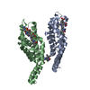
| Entry | Database: PDB / ID: 2vjr | ||||||
|---|---|---|---|---|---|---|---|
| Title | The structure of phycocyanin from Gloeobacter violaceus | ||||||
 Components Components |
| ||||||
 Keywords Keywords |  PHOTOSYNTHESIS / LIGHT HARVESTING PHOTOSYNTHESIS / LIGHT HARVESTING | ||||||
| Function / homology |  Function and homology informationphycobilisome / plasma membrane-derived thylakoid membrane / photosynthesis Function and homology informationphycobilisome / plasma membrane-derived thylakoid membrane / photosynthesisSimilarity search - Function | ||||||
| Biological species |  GLOEOBACTER VIOLACEUS (bacteria) GLOEOBACTER VIOLACEUS (bacteria) | ||||||
| Method | X-RAY DIFFRACTION / SYNCHROTRON / MOLECULAR REPLACEMENT / Resolution: 2.6 Å | ||||||
 Authors Authors | Murray, J.W. / Benson, S. / Nield, J. / Barber, J. | ||||||
 Citation Citation | Journal: To be Published Title: The Structures of the Phycobiliproteins of Gloeobacter Violaceus Authors: Murray, J.W. / Benson, S. / Nield, J. / Barber, J. | ||||||
| History |
|
- Structure visualization
| Structure viewer | Molecule: MolmilJmol/JSmol |
|---|
- Downloads & links
-Download
| PDBx/mmCIF format | 2vjr.cif.gz | 80.8 KB | Display | PDBx/mmCIF format |
|---|---|---|---|---|
| PDB format | pdb2vjr.ent.gz | 60.5 KB | Display | PDB format |
| PDBx/mmJSON format | 2vjr.json.gz | Tree view | PDBx/mmJSON format | |
| Others |  Other downloads Other downloads |
-Validation report
| Arichive directory | https://data.pdbj.org/pub/pdb/validation_reports/vj/2vjrftp://data.pdbj.org/pub/pdb/validation_reports/vj/2vjr | HTTPS FTP |
|---|
-Related structure data
| Related structure data | 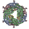 2vjhC 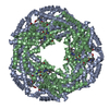 2vjtC 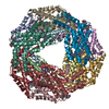 2vmlC 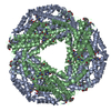 1jboS S: Starting model for refinement C: citing same article ( |
|---|---|
| Similar structure data |


-Links
 PDBj
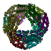
PDBj- Assembly
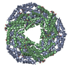
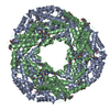
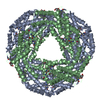
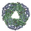
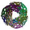
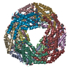
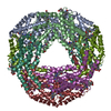
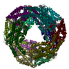
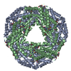
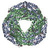
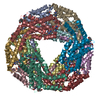
| Deposited unit | 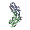
| ||||||||
|---|---|---|---|---|---|---|---|---|---|
| 1 | x 6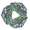
| ||||||||
| Unit cell |
|
-Components
| #1: Protein | Mass: 17679.852 Da / Num. of mol.: 1 / Source method: isolated from a natural source / Details: PASTEUR CULTURE COLLECTION / Source: (natural) GLOEOBACTER VIOLACEUS (bacteria) / Strain: PCC7421 / References: UniProt: Q7M7F7 | ||
|---|---|---|---|
| #2: Protein | Mass: 18478.953 Da / Num. of mol.: 1 / Source method: isolated from a natural source / Details: PASTEUR CULTURE COLLECTION / Source: (natural) GLOEOBACTER VIOLACEUS (bacteria) / Strain: PCC7421 / References: UniProt: Q7M7C7 | ||
| #3: Chemical | Phycocyanobilin  Mass: 588.694 Da / Num. of mol.: 3 / Source method: obtained synthetically / Formula: C33H40N4O6 Mass: 588.694 Da / Num. of mol.: 3 / Source method: obtained synthetically / Formula: C33H40N4O6#4: Water | ChemComp-HOH / | Water Mass: 18.015 Da / Num. of mol.: 84 / Source method: isolated from a natural source / Formula: H2O Mass: 18.015 Da / Num. of mol.: 84 / Source method: isolated from a natural source / Formula: H2O |
-Experimental details
-Experiment
| Experiment | Method: X-RAY DIFFRACTION / Number of used crystals: 1 |
|---|
- Sample preparation
| Crystal | Density Matthews: 3.1 Å3/Da / Density % sol: 60 % / Description: NONE |
|---|---|
| Crystal grow | Method: vapor diffusion, hanging drop Details: 33.3 MM LITHIUM SULFATE, 17 MM TRIS HYDROCHLORIDE PH 8.5, 5% W/V PEG 4K, MIXED WITH AN EQUAL VOLUME OF PROTEIN SOLUTION. HANGING DROP VAPOUR DIFFUSION. |
-Data collection
| Diffraction | Mean temperature: 100 K |
|---|---|
| Diffraction source | Source: SYNCHROTRON / Site: SRS  / Beamline: PX10.1 / Wavelength: 1.283 / Beamline: PX10.1 / Wavelength: 1.283 |
| Detector | Type: MARRESEARCH / Detector: CCD / Date: Nov 8, 2005 |
| Radiation | Protocol: SINGLE WAVELENGTH / Monochromatic (M) / Laue (L): M / Scattering type: x-ray |
| Radiation wavelength | Wavelength: 1.283 Å / Relative weight: 1 |
| Reflection | Resolution: 2.49→29.39 Å / Num. obs: 16967 / % possible obs: 99.6 % / Observed criterion σ(I): 0 / Redundancy: 21.11 % / Rmerge(I) obs: 0.08 / Net I/σ(I): 34.42 |
| Reflection shell | Resolution: 2.49→2.51 Å / Redundancy: 20.96 % / Rmerge(I) obs: 0.41 / Mean I/σ(I) obs: 5.56 / % possible all: 98.8 |
- Processing
| Software |
| ||||||||||||||||||||||||||||||||||||||||||||||||||||||||||||||||||||||||||||||||||||||||||||||||||||||||||||||||||||||||||||||||||||||||||||||||||||||||||||||||||||||||||||||||||||||
|---|---|---|---|---|---|---|---|---|---|---|---|---|---|---|---|---|---|---|---|---|---|---|---|---|---|---|---|---|---|---|---|---|---|---|---|---|---|---|---|---|---|---|---|---|---|---|---|---|---|---|---|---|---|---|---|---|---|---|---|---|---|---|---|---|---|---|---|---|---|---|---|---|---|---|---|---|---|---|---|---|---|---|---|---|---|---|---|---|---|---|---|---|---|---|---|---|---|---|---|---|---|---|---|---|---|---|---|---|---|---|---|---|---|---|---|---|---|---|---|---|---|---|---|---|---|---|---|---|---|---|---|---|---|---|---|---|---|---|---|---|---|---|---|---|---|---|---|---|---|---|---|---|---|---|---|---|---|---|---|---|---|---|---|---|---|---|---|---|---|---|---|---|---|---|---|---|---|---|---|---|---|---|---|
| Refinement | Method to determine structure: MOLECULAR REPLACEMENT Starting model: PDB ENTRY 1JBO Resolution: 2.6→101.53 Å / Cor.coef. Fo:Fc: 0.943 / Cor.coef. Fo:Fc free: 0.913 / SU B: 21.017 / SU ML: 0.218 / TLS residual ADP flag: LIKELY RESIDUAL / Cross valid method: THROUGHOUT / ESU R: 0.5 / ESU R Free: 0.309 / Stereochemistry target values: MAXIMUM LIKELIHOOD / Details: HYDROGENS HAVE BEEN ADDED IN THE RIDING POSITIONS.
| ||||||||||||||||||||||||||||||||||||||||||||||||||||||||||||||||||||||||||||||||||||||||||||||||||||||||||||||||||||||||||||||||||||||||||||||||||||||||||||||||||||||||||||||||||||||
| Solvent computation | Ion probe radii: 0.8 Å / Shrinkage radii: 0.8 Å / VDW probe radii: 1.4 Å / Solvent model: MASK | ||||||||||||||||||||||||||||||||||||||||||||||||||||||||||||||||||||||||||||||||||||||||||||||||||||||||||||||||||||||||||||||||||||||||||||||||||||||||||||||||||||||||||||||||||||||
| Displacement parameters | Biso mean: 28.22 Å2
| ||||||||||||||||||||||||||||||||||||||||||||||||||||||||||||||||||||||||||||||||||||||||||||||||||||||||||||||||||||||||||||||||||||||||||||||||||||||||||||||||||||||||||||||||||||||
| Refinement step | Cycle: LAST / Resolution: 2.6→101.53 Å
| ||||||||||||||||||||||||||||||||||||||||||||||||||||||||||||||||||||||||||||||||||||||||||||||||||||||||||||||||||||||||||||||||||||||||||||||||||||||||||||||||||||||||||||||||||||||
| Refine LS restraints |
|