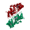
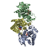
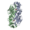
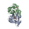
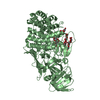
- PDB-2vhd: Crystal Structure Of The Di-Haem Cytochrome C Peroxidase From Pse... -
+
Open data
ID or keywords:
Loading...
-
Basic information
Entry
Database: PDB / ID: 2vhd
Title
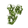
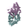
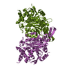
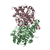
Crystal Structure Of The Di-Haem Cytochrome C Peroxidase From Pseudomonas aeruginosa - Mixed Valence Form
Components
CYTOCHROME C551 PEROXIDASE
Keywords
OXIDOREDUCTASE / IRON / HEME / TRANSPORT / PEROXIDASE / METAL-BINDING / ELECTRON TRANSPORT
Function / homology
Function and homology information
cytochrome-c peroxidase / cytochrome-c peroxidase activity / periplasmic space / electron transfer activity / heme binding / metal ion binding Similarity search - Function
Di-c-type haem protein, MauG/cytochrome c peroxidase / Di-haem cytochrome c peroxidase / Di-haem cytochrome c peroxidase / Cytochrome c / Cytochrome c-like domain / Cytochrome Bc1 Complex; Chain D, domain 2 / Cytochrome c family profile. / Cytochrome c-like domain / Cytochrome c-like domain superfamily / Orthogonal Bundle / Mainly Alpha Similarity search - Domain/homology
 Movie
Movie Controller

 Yorodumi
 Open data
 Basic information
 Components
 Keywords
 OXIDOREDUCTASE /
  Function and homology information

 Authors
 Citation
 Structure visualization
 Downloads & links
 Other downloads
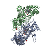


 PDBj
PDBj


 Assembly
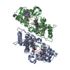

 Mass: 618.503 Da / Num. of mol.: 4 / Source method: obtained synthetically / Formula: C34H34FeN4O4
 Mass: 40.078 Da / Num. of mol.: 2 / Source method: obtained synthetically / Formula: Ca
 Mass: 18.015 Da / Num. of mol.: 288 / Source method: isolated from a natural source / Formula: H2O
 Sample preparation
 / Beamline: X11 / Wavelength: 0.8115
  Processing