 Movie
Movie Controller

[English] 日本語
 Yorodumi
- PDB-5xc2: Crystal structure of GH family 81 beta-1,3-glucanase from Rhizomu... -
+ Open data

- Basic information
| Entry | Database: PDB / ID: 5xc2 | ||||||||||||
|---|---|---|---|---|---|---|---|---|---|---|---|---|---|
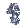
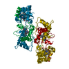
| Title | Crystal structure of GH family 81 beta-1,3-glucanase from Rhizomucr miehei complexed with laminarihexaose | ||||||||||||
 Components Components | Endo-beta-1,3-glucanase | ||||||||||||
 Keywords Keywords |  HYDROLASE / glycoside hydrolase family 81 / catalytic mechanism HYDROLASE / glycoside hydrolase family 81 / catalytic mechanism | ||||||||||||
| Function / homology |  Function and homology information Function and homology informationglucan endo-1,4-beta-glucanase activity, C-3 substituted reducing group / glucan endo-1,3-beta-glucanase activity, C-3 substituted reducing group / glucan endo-1,3-beta-D-glucosidase / glucan endo-1,3-beta-D-glucosidase activity / polysaccharide catabolic process / cell wall organization / extracellular region Similarity search - Function | ||||||||||||
| Biological species |  Rhizomucor miehei (fungus) Rhizomucor miehei (fungus) | ||||||||||||
| Method | X-RAY DIFFRACTION / SYNCHROTRON / MOLECULAR REPLACEMENT / Resolution: 2.7 Å | ||||||||||||
 Authors Authors | Qin, Z. / Yang, S. / Peng, Z. / Yan, Q. / Jiang, Z. | ||||||||||||
 Citation Citation | Journal: To Be Published Title: Catalytic mechanism of glycoside hydrolase family 81 beta-1,3-glucanase Authors: Yang, S. / Qin, Z. / Zhou, P. / Yan, Q. / Jiang, Z. | ||||||||||||
| History |
|
- Structure visualization
| Structure viewer | Molecule: MolmilJmol/JSmol |
|---|
- Downloads & links
-Download
| PDBx/mmCIF format | 5xc2.cif.gz | 568.3 KB | Display | PDBx/mmCIF format |
|---|---|---|---|---|
| PDB format | pdb5xc2.ent.gz | 468.7 KB | Display | PDB format |
| PDBx/mmJSON format | 5xc2.json.gz | Tree view | PDBx/mmJSON format | |
| Others |  Other downloads Other downloads |
-Validation report
| Arichive directory | https://data.pdbj.org/pub/pdb/validation_reports/xc/5xc2ftp://data.pdbj.org/pub/pdb/validation_reports/xc/5xc2 | HTTPS FTP |
|---|
-Related structure data
| Related structure data | 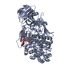 5xbzC 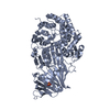 4k3aS C: citing same article ( S: Starting model for refinement |
|---|---|
| Similar structure data |


-Links
 PDBj
PDBj- Assembly
| Deposited unit | 
| ||||||||
|---|---|---|---|---|---|---|---|---|---|
| 1 | 
| ||||||||
| 2 | 
| ||||||||
| 3 | 
| ||||||||
| 4 | 
| ||||||||
| Unit cell |
|
-Components
-Protein / Non-polymers , 2 types, 483 molecules ABCD
| #1: Protein | Mass: 89586.039 Da / Num. of mol.: 4 Source method: isolated from a genetically manipulated source Source: (gene. exp.) Rhizomucor miehei (fungus)Production host:  Escherichia coli 'BL21-Gold(DE3)pLysS AG' (bacteria) Escherichia coli 'BL21-Gold(DE3)pLysS AG' (bacteria)References: UniProt: A0A023I7E1, glucan endo-1,3-beta-D-glucosidase #7: Water | ChemComp-HOH / | WaterMass: 18.015 Da / Num. of mol.: 479 / Source method: isolated from a natural source / Formula: H2O |
|---|
-Sugars , 5 types, 14 molecules
| #2: Polysaccharide | beta-D-glucopyranose-(1-3)-beta-D-glucopyranose-(1-3)-beta-D-glucopyranose-(1-3)-beta-D- ...beta-D-glucopyranose-(1-3)-beta-D-glucopyranose-(1-3)-beta-D-glucopyranose-(1-3)-beta-D-glucopyranose-(1-3)-beta-D-glucopyranose-(1-3)-beta-D-glucopyranose / Mass: 990.860 Da / Num. of mol.: 1 Source method: isolated from a genetically manipulated source | ||||||
|---|---|---|---|---|---|---|---|
| #3: Polysaccharide | / Mass: 828.719 Da / Num. of mol.: 2 Source method: isolated from a genetically manipulated source #4: Polysaccharide | beta-D-glucopyranose-(1-3)-beta-D-glucopyranose-(1-3)-beta-D-glucopyranose-(1-3)-beta-D-glucopyranose / Mass: 666.578 Da / Num. of mol.: 5Source method: isolated from a genetically manipulated source #5: Polysaccharide | beta-D-glucopyranose-(1-3)-beta-D-glucopyranose-(1-3)-beta-D-glucopyranose / Mass: 504.438 Da / Num. of mol.: 4Source method: isolated from a genetically manipulated source #6: Polysaccharide |   , Oligosaccharide / Class: Antimicrobial / Mass: 342.297 Da / Num. of mol.: 2 , Oligosaccharide / Class: Antimicrobial / Mass: 342.297 Da / Num. of mol.: 2Source method: isolated from a genetically manipulated source Details: oligosaccharide / References: beta-laminaribiose |
-Experimental details
-Experiment
| Experiment | Method: X-RAY DIFFRACTION / Number of used crystals: 1 |
|---|
- Sample preparation
| Crystal | Density Matthews: 1.94 Å3/Da / Density % sol: 36.48 % |
|---|---|
| Crystal grow | Temperature: 293 K / Method: vapor diffusion, sitting drop Details: laminarihexaose (G6) was added to the protein solution at a final concentration of 1%(w/v). The complex crystals were obtained in a reservoir solution containing 24%(w/v) PEG4000, 80 mM Tris- ...Details: laminarihexaose (G6) was added to the protein solution at a final concentration of 1%(w/v). The complex crystals were obtained in a reservoir solution containing 24%(w/v) PEG4000, 80 mM Tris-HCl (pH 8.5), and 6%(v/v) MPD |
-Data collection
| Diffraction | Mean temperature: 100 K |
|---|---|
| Diffraction source | Source: SYNCHROTRON / Site: Photon Factory  / Beamline: BL-1A / Wavelength: 1.1 Å / Beamline: BL-1A / Wavelength: 1.1 Å |
| Detector | Type: MAR CCD 130 mm / Detector: CCD / Date: Dec 12, 2014 |
| Radiation | Protocol: SINGLE WAVELENGTH / Monochromatic (M) / Laue (L): M / Scattering type: x-ray |
| Radiation wavelength | Wavelength: 1.1 Å / Relative weight: 1 |
| Reflection | Resolution: 2.7→50 Å / Num. obs: 74950 / % possible obs: 99.55 % / Redundancy: 17 % / Rmerge(I) obs: 0.102 / Net I/σ(I): 10.37 |
| Reflection shell | Resolution: 2.7→2.94 Å / Rmerge(I) obs: 0.638 / Mean I/σ(I) obs: 2.36 / % possible all: 99.16 |
- Processing
| Software |
| |||||||||||||||||||||||||||||||||||||||||||||||||||||||||||||||||||||||||||||||||||||||||||||||||||||||||
|---|---|---|---|---|---|---|---|---|---|---|---|---|---|---|---|---|---|---|---|---|---|---|---|---|---|---|---|---|---|---|---|---|---|---|---|---|---|---|---|---|---|---|---|---|---|---|---|---|---|---|---|---|---|---|---|---|---|---|---|---|---|---|---|---|---|---|---|---|---|---|---|---|---|---|---|---|---|---|---|---|---|---|---|---|---|---|---|---|---|---|---|---|---|---|---|---|---|---|---|---|---|---|---|---|---|---|
| Refinement | Method to determine structure: MOLECULAR REPLACEMENT Starting model: 4k3a Resolution: 2.7→29.73 Å / SU ML: 0.36 / Cross valid method: FREE R-VALUE / σ(F): 1.36 / Phase error: 23.44 / Stereochemistry target values: ML
| |||||||||||||||||||||||||||||||||||||||||||||||||||||||||||||||||||||||||||||||||||||||||||||||||||||||||
| Solvent computation | Shrinkage radii: 0.9 Å / VDW probe radii: 1.11 Å / Solvent model: FLAT BULK SOLVENT MODEL | |||||||||||||||||||||||||||||||||||||||||||||||||||||||||||||||||||||||||||||||||||||||||||||||||||||||||
| Refinement step | Cycle: LAST / Resolution: 2.7→29.73 Å
| |||||||||||||||||||||||||||||||||||||||||||||||||||||||||||||||||||||||||||||||||||||||||||||||||||||||||
| Refine LS restraints |
| |||||||||||||||||||||||||||||||||||||||||||||||||||||||||||||||||||||||||||||||||||||||||||||||||||||||||
| LS refinement shell |
|
