 Movie
Movie Controller

[English] 日本語
 Yorodumi
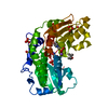
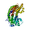
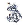
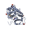
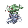
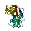
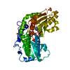
- PDB-2vf5: Glucosamine-6-phosphate synthase in complex with glucosamine-6- p... -
+ Open data

- Basic information
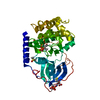
| Entry | Database: PDB / ID: 2vf5 | ||||||
|---|---|---|---|---|---|---|---|
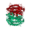
| Title | Glucosamine-6-phosphate synthase in complex with glucosamine-6- phosphate | ||||||
 Components Components | GLUCOSAMINE--FRUCTOSE-6-PHOSPHATE AMINOTRANSFERASE | ||||||
 Keywords Keywords |  TRANSFERASE / GLUCOSAMINE-6- PHOSPHATE SYNTHASE / N TERMINAL NUCLEOPHILE / GLUTAMINE AMIDOTRANSFERASE / AMIDOTRANSFERASE / AMMONIA-CHANNELING / AMINOTRANSFERASE TRANSFERASE / GLUCOSAMINE-6- PHOSPHATE SYNTHASE / N TERMINAL NUCLEOPHILE / GLUTAMINE AMIDOTRANSFERASE / AMIDOTRANSFERASE / AMMONIA-CHANNELING / AMINOTRANSFERASE | ||||||
| Function / homology |  Function and homology informationglutamine-fructose-6-phosphate transaminase (isomerizing) / glutamine-fructose-6-phosphate transaminase (isomerizing) activity / UDP-N-acetylglucosamine metabolic process / UDP-N-acetylglucosamine biosynthetic process / carbohydrate derivative binding / fructose 6-phosphate metabolic process / protein N-linked glycosylation / glutamine metabolic process / carbohydrate metabolic process / cytosol Function and homology informationglutamine-fructose-6-phosphate transaminase (isomerizing) / glutamine-fructose-6-phosphate transaminase (isomerizing) activity / UDP-N-acetylglucosamine metabolic process / UDP-N-acetylglucosamine biosynthetic process / carbohydrate derivative binding / fructose 6-phosphate metabolic process / protein N-linked glycosylation / glutamine metabolic process / carbohydrate metabolic process / cytosolSimilarity search - Function | ||||||
| Biological species |  ESCHERICHIA COLI (E. coli) ESCHERICHIA COLI (E. coli) | ||||||
| Method | X-RAY DIFFRACTION / SYNCHROTRON / MOLECULAR REPLACEMENT / Resolution: 2.9 Å | ||||||
 Authors Authors | Mouilleron, S. / Golinelli-Pimpaneau, B. | ||||||
 Citation Citation | Journal: J.Mol.Biol. / Year: 2008 Title: Ordering of C-Terminal Loop and Glutaminase Domains of Glucosamine-6-Phosphate Synthase Promotes Sugar Ring Opening and Formation of the Ammonia Channel. Authors: Mouilleron, S. / Badet-Denisot, M.-A. / Golinelli-Pimpaneau, B. #1: Journal: J.Biol.Chem. / Year: 2006Title: Glutamine Binding Opens the Ammonia Channel and Activates Glucosamine-6-P Synthase. Authors: Mouilleron, S. / Badet-Denisot, M.-A. / Golinelli-Pimpaneau, B. | ||||||
| History |
|
- Structure visualization
| Structure viewer | Molecule: MolmilJmol/JSmol |
|---|
- Downloads & links
-Download
| PDBx/mmCIF format | 2vf5.cif.gz | 87.4 KB | Display | PDBx/mmCIF format |
|---|---|---|---|---|
| PDB format | pdb2vf5.ent.gz | 63.9 KB | Display | PDB format |
| PDBx/mmJSON format | 2vf5.json.gz | Tree view | PDBx/mmJSON format | |
| Others |  Other downloads Other downloads |
-Validation report
| Arichive directory | https://data.pdbj.org/pub/pdb/validation_reports/vf/2vf5ftp://data.pdbj.org/pub/pdb/validation_reports/vf/2vf5 | HTTPS FTP |
|---|
-Related structure data
| Related structure data |  2vf4C 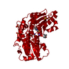 1moqS S: Starting model for refinement C: citing same article ( |
|---|---|
| Similar structure data |


-Links
 PDBj
PDBj


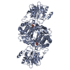
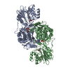
- Assembly
| Deposited unit | 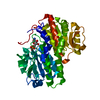
| ||||||||
|---|---|---|---|---|---|---|---|---|---|
| 1 |
| ||||||||
| Unit cell |
|
-Components
| #1: Protein | Mass: 66846.016 Da / Num. of mol.: 1 / Fragment: RESIDUES 2-609 Source method: isolated from a genetically manipulated source Source: (gene. exp.) ESCHERICHIA COLI (E. coli) / Plasmid: PMA1 / Production host: ESCHERICHIA COLI (E. coli) / Strain (production host): HB101References: UniProt: P17169, glutamine-fructose-6-phosphate transaminase (isomerizing) |
|---|---|
| #2: Sugar | ChemComp-GLP / 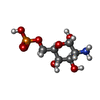  Type: D-saccharide, alpha linking / Mass: 259.151 Da / Num. of mol.: 1 Type: D-saccharide, alpha linking / Mass: 259.151 Da / Num. of mol.: 1Source method: isolated from a genetically manipulated source Formula: C6H14NO8P |
| #3: Water | ChemComp-HOH / Water Mass: 18.015 Da / Num. of mol.: 12 / Source method: isolated from a natural source / Formula: H2O Mass: 18.015 Da / Num. of mol.: 12 / Source method: isolated from a natural source / Formula: H2O |
-Experimental details
-Experiment
| Experiment | Method: X-RAY DIFFRACTION / Number of used crystals: 1 |
|---|
- Sample preparation
| Crystal | Density Matthews: 2.58 Å3/Da / Density % sol: 52.36 % / Description: NONE |
|---|---|
| Crystal grow | pH: 5.5 / Details: 8% PEG4000, 0.2 M SODIUM ACETATE PH 5.5 |
-Data collection
| Diffraction | Mean temperature: 100 K |
|---|---|
| Diffraction source | Source: SYNCHROTRON / Site: ESRF  / Beamline: ID23-1 / Wavelength: 1.07225 / Beamline: ID23-1 / Wavelength: 1.07225 |
| Detector | Type: ADSC CCD / Detector: CCD / Date: Jul 21, 2006 |
| Radiation | Protocol: SINGLE WAVELENGTH / Monochromatic (M) / Laue (L): M / Scattering type: x-ray |
| Radiation wavelength | Wavelength: 1.07225 Å / Relative weight: 1 |
| Reflection | Resolution: 2.9→20 Å / Num. obs: 15522 / % possible obs: 99.2 % / Observed criterion σ(I): 0 / Redundancy: 4.6 % / Rmerge(I) obs: 0.08 / Net I/σ(I): 5.3 |
| Reflection shell | Resolution: 2.9→3.06 Å / Redundancy: 4.7 % / Rmerge(I) obs: 0.51 / Mean I/σ(I) obs: 1.5 / % possible all: 99.2 |
- Processing
| Software |
| ||||||||||||||||||||||||||||||||||||||||||||||||||||||||||||||||||||||||||||||||||||||||||||||||||||||||||||||||||||||||||||||||||||||||||||||||||||||||||||||||||||||||||||||||||||||
|---|---|---|---|---|---|---|---|---|---|---|---|---|---|---|---|---|---|---|---|---|---|---|---|---|---|---|---|---|---|---|---|---|---|---|---|---|---|---|---|---|---|---|---|---|---|---|---|---|---|---|---|---|---|---|---|---|---|---|---|---|---|---|---|---|---|---|---|---|---|---|---|---|---|---|---|---|---|---|---|---|---|---|---|---|---|---|---|---|---|---|---|---|---|---|---|---|---|---|---|---|---|---|---|---|---|---|---|---|---|---|---|---|---|---|---|---|---|---|---|---|---|---|---|---|---|---|---|---|---|---|---|---|---|---|---|---|---|---|---|---|---|---|---|---|---|---|---|---|---|---|---|---|---|---|---|---|---|---|---|---|---|---|---|---|---|---|---|---|---|---|---|---|---|---|---|---|---|---|---|---|---|---|---|
| Refinement | Method to determine structure: MOLECULAR REPLACEMENT Starting model: PDB ENTRY 1MOQ Resolution: 2.9→15 Å / Cor.coef. Fo:Fc: 0.947 / Cor.coef. Fo:Fc free: 0.934 / SU B: 12.78 / SU ML: 0.24 / TLS residual ADP flag: LIKELY RESIDUAL / Cross valid method: THROUGHOUT / ESU R: 0.608 / ESU R Free: 0.316 / Stereochemistry target values: MAXIMUM LIKELIHOOD Details: HYDROGENS HAVE BEEN ADDED IN THE RIDING POSITIONS. THE GLUTAMINASE DOMAIN THAT WAS DISORDERED WAS NOT MODELLED.
| ||||||||||||||||||||||||||||||||||||||||||||||||||||||||||||||||||||||||||||||||||||||||||||||||||||||||||||||||||||||||||||||||||||||||||||||||||||||||||||||||||||||||||||||||||||||
| Solvent computation | Ion probe radii: 0.8 Å / Shrinkage radii: 0.8 Å / VDW probe radii: 1.4 Å / Solvent model: BABINET MODEL WITH MASK | ||||||||||||||||||||||||||||||||||||||||||||||||||||||||||||||||||||||||||||||||||||||||||||||||||||||||||||||||||||||||||||||||||||||||||||||||||||||||||||||||||||||||||||||||||||||
| Displacement parameters | Biso mean: 27.46 Å2
| ||||||||||||||||||||||||||||||||||||||||||||||||||||||||||||||||||||||||||||||||||||||||||||||||||||||||||||||||||||||||||||||||||||||||||||||||||||||||||||||||||||||||||||||||||||||
| Refinement step | Cycle: LAST / Resolution: 2.9→15 Å
| ||||||||||||||||||||||||||||||||||||||||||||||||||||||||||||||||||||||||||||||||||||||||||||||||||||||||||||||||||||||||||||||||||||||||||||||||||||||||||||||||||||||||||||||||||||||
| Refine LS restraints |
|