 Movie
Movie Controller

[English] 日本語
 Yorodumi
- PDB-1xfg: Glutaminase domain of glucosamine 6-phosphate synthase complexed ... -
+ Open data

- Basic information
| Entry | Database: PDB / ID: 1xfg | |||||||||
|---|---|---|---|---|---|---|---|---|---|---|
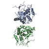
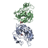
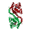
| Title | Glutaminase domain of glucosamine 6-phosphate synthase complexed with l-glu hydroxamate | |||||||||
 Components Components | Glucosamine--fructose-6-phosphate aminotransferase [isomerizing] | |||||||||
 Keywords Keywords |  TRANSFERASE / GLUTAMINE AMIDOTRANSFERASE / N-TERMINAL NUCLEOPHILE TRANSFERASE / GLUTAMINE AMIDOTRANSFERASE / N-TERMINAL NUCLEOPHILE | |||||||||
| Function / homology |  Function and homology informationglutamine-fructose-6-phosphate transaminase (isomerizing) / glutamine-fructose-6-phosphate transaminase (isomerizing) activity / UDP-N-acetylglucosamine metabolic process / UDP-N-acetylglucosamine biosynthetic process / carbohydrate derivative binding / fructose 6-phosphate metabolic process / protein N-linked glycosylation / glutamine metabolic process / carbohydrate metabolic process / cytosol Function and homology informationglutamine-fructose-6-phosphate transaminase (isomerizing) / glutamine-fructose-6-phosphate transaminase (isomerizing) activity / UDP-N-acetylglucosamine metabolic process / UDP-N-acetylglucosamine biosynthetic process / carbohydrate derivative binding / fructose 6-phosphate metabolic process / protein N-linked glycosylation / glutamine metabolic process / carbohydrate metabolic process / cytosolSimilarity search - Function | |||||||||
| Biological species |  Escherichia coli (E. coli) Escherichia coli (E. coli) | |||||||||
| Method | X-RAY DIFFRACTION / SYNCHROTRON / MOLECULAR REPLACEMENT / Resolution: 1.85 Å | |||||||||
 Authors Authors | Isupov, M.N. / Teplyakov, A. | |||||||||
 Citation Citation | Journal: Structure / Year: 1996 Title: Substrate Binding is Required for Assembly of the Active Conformation of the Catalytic Site in Ntn Amidotransferases: Evidence from the 1.8 Angstrom Crystal Structure of the Glutaminase Domain ...Title: Substrate Binding is Required for Assembly of the Active Conformation of the Catalytic Site in Ntn Amidotransferases: Evidence from the 1.8 Angstrom Crystal Structure of the Glutaminase Domain of Glucosamine 6-Phosphate Synthase Authors: Isupov, M.N. / Obmolova, G. / Butterworth, S. / Badet-Denisot, M.-A. / Badet, B. / Polikarpov, I. / Littlechild, J.A. / Teplyakov, A. #1: Journal: J.Mol.Biol. / Year: 1994Title: Crystallization and Preliminary X-Ray Analysis of the Two Domains of Glucosamine-6-Phosphate Synthase from Escherichia Coli Authors: Obmolova, G. / Badet-Denisot, M.A. / Badet, B. / Teplyakov, A. #2: Journal: J.Mol.Biol. / Year: 2001Title: Channeling of Ammonia in Glucosamine 6-Phosphate Synthase Authors: Teplyakov, A. / Obmolova, G. / Badet-Denisot, M.A. / Badet, B. | |||||||||
| History |
|
- Structure visualization
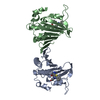
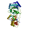
| Structure viewer | Molecule: MolmilJmol/JSmol |
|---|
- Downloads & links
-Download
| PDBx/mmCIF format | 1xfg.cif.gz | 113.7 KB | Display | PDBx/mmCIF format |
|---|---|---|---|---|
| PDB format | pdb1xfg.ent.gz | 86.8 KB | Display | PDB format |
| PDBx/mmJSON format | 1xfg.json.gz | Tree view | PDBx/mmJSON format | |
| Others |  Other downloads Other downloads |
-Validation report
| Arichive directory | https://data.pdbj.org/pub/pdb/validation_reports/xf/1xfgftp://data.pdbj.org/pub/pdb/validation_reports/xf/1xfg | HTTPS FTP |
|---|
-Related structure data
| Related structure data |  1xffC 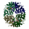 1gphS C: citing same article ( S: Starting model for refinement |
|---|---|
| Similar structure data |

-Links
 PDBj
PDBj


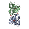
- Assembly
| Deposited unit | 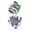
| ||||||||||
|---|---|---|---|---|---|---|---|---|---|---|---|
| 1 |
| ||||||||||
| Unit cell |
|
-Components
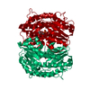
| #1: Protein | Mass: 26507.059 Da / Num. of mol.: 2 / Fragment: GLUTAMINASE DOMAIN Source method: isolated from a genetically manipulated source Details: inhibited by L-GLUTAMATE / Source: (gene. exp.) Escherichia coli (E. coli) / Strain: 3000HFR / Gene: GLMS / Plasmid: PGM10A / Production host: Escherichia coli (E. coli)References: UniProt: P17169, glutamine-fructose-6-phosphate transaminase (isomerizing)#2: Chemical |   Mass: 22.990 Da / Num. of mol.: 2 / Source method: obtained synthetically / Formula: Na Mass: 22.990 Da / Num. of mol.: 2 / Source method: obtained synthetically / Formula: Na#3: Chemical | ChemComp-ACT / | Acetate  Mass: 59.044 Da / Num. of mol.: 1 / Source method: obtained synthetically / Formula: C2H3O2 Mass: 59.044 Da / Num. of mol.: 1 / Source method: obtained synthetically / Formula: C2H3O2#4: Chemical | 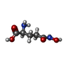  Mass: 162.144 Da / Num. of mol.: 2 / Source method: obtained synthetically / Formula: C5H10N2O4 Mass: 162.144 Da / Num. of mol.: 2 / Source method: obtained synthetically / Formula: C5H10N2O4#5: Water | ChemComp-HOH / | Water Mass: 18.015 Da / Num. of mol.: 295 / Source method: isolated from a natural source / Formula: H2O Mass: 18.015 Da / Num. of mol.: 295 / Source method: isolated from a natural source / Formula: H2O |
|---|
-Experimental details
-Experiment
| Experiment | Method: X-RAY DIFFRACTION / Number of used crystals: 1 |
|---|
- Sample preparation
| Crystal | Density Matthews: 2.34 Å3/Da / Density % sol: 47.3 % |
|---|---|
| Crystal grow | Temperature: 295 K / Method: vapor diffusion, hanging drop / pH: 6.5 Details: 0.1 M cacodylate, 1 M sodium acetate, 20% PEG 4000, pH 6.5, VAPOR DIFFUSION, HANGING DROP, temperature 295K |
-Data collection
| Diffraction | Mean temperature: 295 K |
|---|---|
| Diffraction source | Source: SYNCHROTRON / Site: EMBL/DESY, HAMBURG  / Beamline: X11 / Wavelength: 0.93 / Wavelength: 0.93 Å / Beamline: X11 / Wavelength: 0.93 / Wavelength: 0.93 Å |
| Detector | Type: MARRESEARCH / Detector: IMAGE PLATE / Date: Nov 18, 1993 / Details: MIRROR |
| Radiation | Monochromator: GRAPHITE / Protocol: SINGLE WAVELENGTH / Monochromatic (M) / Laue (L): M / Scattering type: x-ray |
| Radiation wavelength | Wavelength: 0.93 Å / Relative weight: 1 |
| Reflection | Resolution: 1.85→15 Å / Num. all: 42930 / Num. obs: 42930 / % possible obs: 99.6 % / Observed criterion σ(I): -3 / Redundancy: 4.4 % / Biso Wilson estimate: 18.4 Å2 / Rmerge(I) obs: 0.053 / Net I/σ(I): 15.2 |
| Reflection shell | Resolution: 1.85→1.9 Å / Redundancy: 4 % / Rmerge(I) obs: 0.262 / Mean I/σ(I) obs: 3.2 / % possible all: 92.3 |
- Processing
| Software |
| ||||||||||||||||||||||||||||||||||||||||||||||||||||||||||||||||||||||||||||||||
|---|---|---|---|---|---|---|---|---|---|---|---|---|---|---|---|---|---|---|---|---|---|---|---|---|---|---|---|---|---|---|---|---|---|---|---|---|---|---|---|---|---|---|---|---|---|---|---|---|---|---|---|---|---|---|---|---|---|---|---|---|---|---|---|---|---|---|---|---|---|---|---|---|---|---|---|---|---|---|---|---|---|
| Refinement | Method to determine structure: MOLECULAR REPLACEMENT Starting model: 1GPH Resolution: 1.85→15 Å / Cor.coef. Fo:Fc: 0.974 / SU B: 1.829 / SU ML: 0.056 / Cross valid method: none used / σ(F): 0 / ESU R: 0.114
| ||||||||||||||||||||||||||||||||||||||||||||||||||||||||||||||||||||||||||||||||
| Solvent computation | Ion probe radii: 0.8 Å / Shrinkage radii: 0.8 Å / VDW probe radii: 1.2 Å / Solvent model: BABINET MODEL WITH MASK | ||||||||||||||||||||||||||||||||||||||||||||||||||||||||||||||||||||||||||||||||
| Displacement parameters | Biso mean: 26.6 Å2
| ||||||||||||||||||||||||||||||||||||||||||||||||||||||||||||||||||||||||||||||||
| Refine analyze | Luzzati coordinate error obs: 0.114 Å / Luzzati sigma a obs: 0.056 Å | ||||||||||||||||||||||||||||||||||||||||||||||||||||||||||||||||||||||||||||||||
| Refinement step | Cycle: LAST / Resolution: 1.85→15 Å
| ||||||||||||||||||||||||||||||||||||||||||||||||||||||||||||||||||||||||||||||||
| Refine LS restraints |
| ||||||||||||||||||||||||||||||||||||||||||||||||||||||||||||||||||||||||||||||||
| LS refinement shell | Resolution: 1.85→1.9 Å / Total num. of bins used: 20
|
