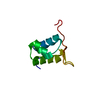
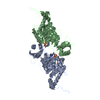
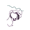
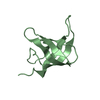
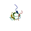
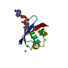
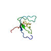
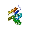
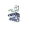
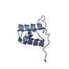
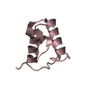
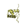
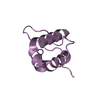
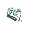
A: DNA TRANSLOCASE FTSK B: DNA TRANSLOCASE FTSK C: DNA TRANSLOCASE FTSK D: DNA TRANSLOCASE FTSK E: DNA TRANSLOCASE FTSK F: DNA TRANSLOCASE FTSK G: DNA TRANSLOCASE FTSK H: DNA TRANSLOCASE FTSK
Resolution: 1.4→95.35 Å / Cor.coef. Fo:Fc: 0.968 / Cor.coef. Fo:Fc free: 0.955 / SU B: 1.963 / SU ML: 0.036 / Cross valid method: THROUGHOUT / ESU R: 0.063 / ESU R Free: 0.062 / Stereochemistry target values: MAXIMUM LIKELIHOOD / Details: HYDROGENS HAVE BEEN ADDED IN THE RIDING POSITIONS.
Rfactor
Num. reflection
% reflection
Selection details
Rfree
0.192
4842
5 %
RANDOM
Rwork
0.144
-
-
-
obs
0.146
91374
99.8 %
-
Solvent computation
Ion probe radii: 0.8 Å / Shrinkage radii: 0.8 Å / VDW probe radii: 1.4 Å / Solvent model: BABINET MODEL WITH MASK
 Movie
Movie Controller


 Open data
 Basic information
 Components
 Keywords
 TRANSPORT PROTEIN / NUCLEOTIDE-BINDING / CHROMOSOME PARTITION / ATP-BINDING / DNA-BINDING /
  Function and homology information

 Authors
 Citation
 Structure visualization
 Downloads & links
 Other downloads
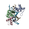


 PDBj
PDBj Assembly
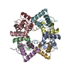


 Mass: 18.015 Da / Num. of mol.: 661 / Source method: isolated from a natural source / Formula: H2O
 Sample preparation
 / Beamline: ID14-4 / Wavelength: 0.9393
  Processing